Japanese
English
- 有料閲覧
- Abstract 文献概要
- 1ページ目 Look Inside
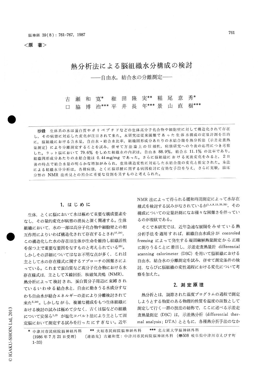
抄録 生体系の水は蛋白質やポリペプチドなどの生体高分子化合物や細胞壁に対して構造化されて存在し,その病態に対応した変化が注目されて来た。本研究は従来困難であった生体水構成の定量計測を目的に,脳組織における含水量,自由水・結合水比率,組織固形成分あたりの水結合能を熱分析法(示差走査熱量測定)により分離測定することを試み,併せて方法論上の信頼性,病態研究への今後の応用につき考察した。ラット脳において79.6%をしめた組織水の内訳は,自由水88.9%,結合水11.1%の比率であり,組織固形成分あたりの水結合能は0.44mg/mgであった。さらに脳組織における死後変化をみると,2日後の時点で結合水量の明らかな増加がみられ,生体構造変性に対応した水結合能の変化と推定された。本法による組織水分分析は,各種病態,とくに脳浮腫に関する病因検討に有効な手段を与え,さらに実験,臨床分野のNMR法所見との照合に重要な役割を果すものと考えられた。
In the living system, tissue water is considered to be composed of both free and bound water. Bound water encompasses the structural water of the cell wall and of various biological substances of high molecular weight, such as proteins and polypeptides. The present study was designed to measure thermoanalytically free and bound water on a quantitative basis in fresh brain of rats using differential scanning calorimetry (DSC).
Our intention was to determine the fraction of freezable water in tissue. Freezable water in a tissue represents the fraction of free water. In the present study, freezing was conducted at a con-stant rate of -10℃/min from room temperature to-75℃ by a SSC/560 S (Seiko Electronics). The system allows for calculation of the amount of free water from the differential scanning calorimetry curve employing a coloric constant of 79.4 cal/mg. Aluminium oxide was used as calorimetric refer-ence. The fraction of bound water was calculated by subtraction of the amount of free water from that of total tissue water. Water binding to solid tissue component was estimated from tissue dry weight and the bound water fraction.
Mean water content of normal gray matter in adult Wistar rats was 76.9±1.4% (SD). 88.9% of total tissue water was free whereas 11.1±2.8% (SD) was bound. Bound water of brain parenchyma amounted to 0.44±0.12 mg/mg dry weight. As compared to other tissues such as cardiac muscle and liver, brain parenchyma obviously exceeded in free water content. The total water content of serum was 94.4±1.2%; 90.7±2.6% was free and 9.3±2.6% (SD) was bound. The amount of bound water in serum was around fourfold, being 1.61± 0.45 (SD) mg/mg dry weight. This seemed to be a proper compositional feature of blood, which exists separately from parenchymal tissue by vessels.
From the observation on postmortem changes in tissue water status, it was of evidence that water binding to brain tissue component markedly in-creased at the period of 48 hours after sacrifice. This probably corresponds to the conformation changes occured in brain parenchyma.
The present trials for quantitative determination of free and bound water components in brain tis-sue can be applied for analysis of tissue water status in combination with various pathophysio-logies. In particular, the technology mentioned pos-sibly plays an important role to clarify the back ground of dynamics of brain edema. Furthermore, those quantitative data on tissue water are con-vinced to contribute to profound understanding of findings in nuclear magnetic resonance imagings and proton relaxation time studies.

Copyright © 1987, Igaku-Shoin Ltd. All rights reserved.


