Japanese
English
- 有料閲覧
PPV購入案内
1,320円 (1,200円+税10%) こちらは、635ページから640ページの文献です。購入には医書.jp本会員登録が必要です。
会員登録完了後に改めて上記「購入サイトへ」を選択してください。
または「購入サイトへ」選択後に「購入ログイン」、「新規会員登録」の順に進んでください。
会員登録について詳しくはをご確認ください。
- Abstract 文献概要
- 1ページ目 Look Inside
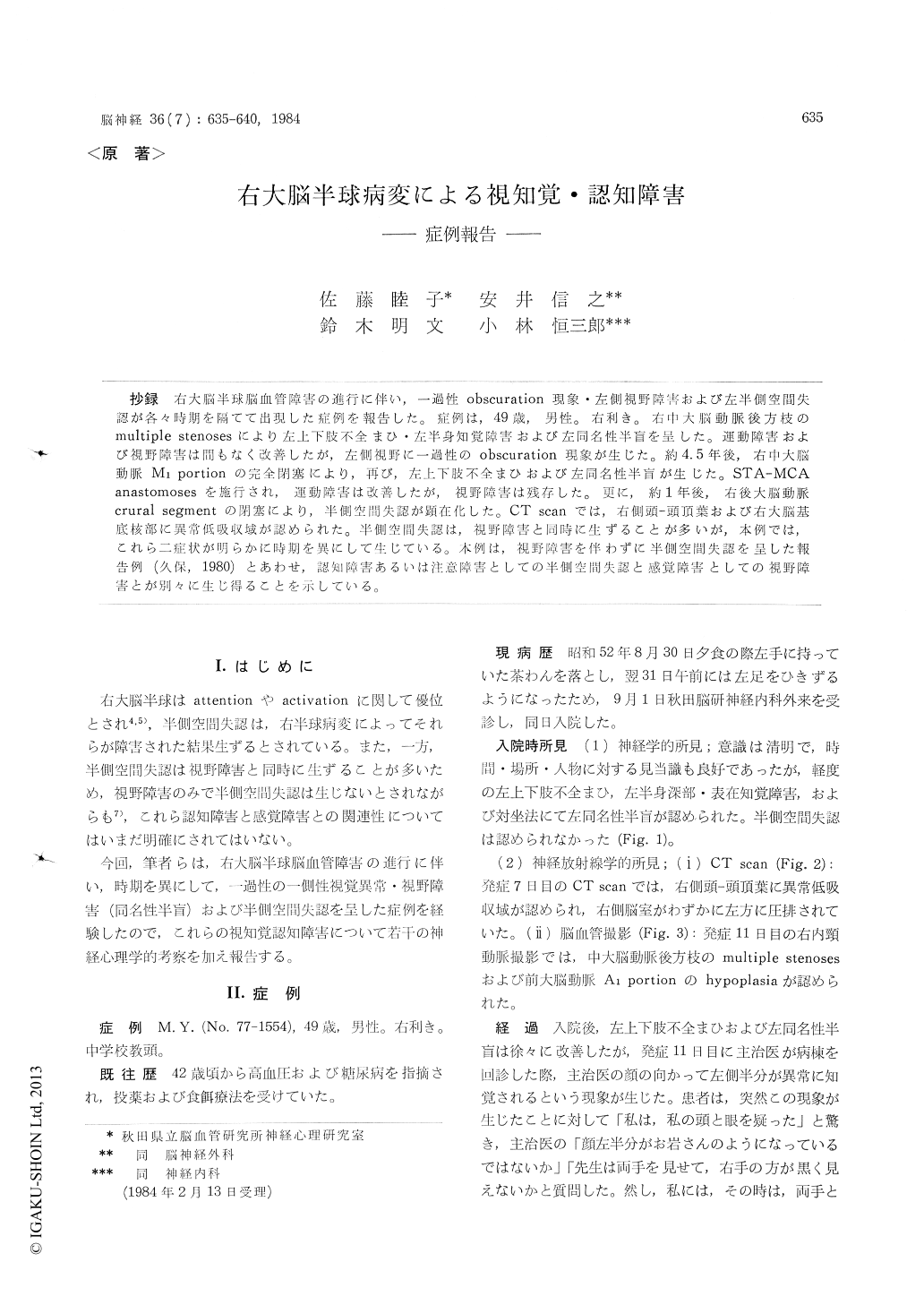
抄録 右大脳半球脳血管障害の進行に伴い,一過性obscuration現象・左側視野障害および左半側空間失認が各々時期を隔てて出現した症例を報告した。症例は,49歳,男性。右利き。右中大脳動脈後方枝のmultiple stenosesにより左上下肢不全まひ・左半身知覚障害および左同名性半盲を呈した。運動障害および視野障害は間もなく改善したが,左側視野に一過性のobscuration現象が生じた。約4.5年後,右中大脳動脈M1portionの完全閉塞により,再び,左上下肢不全まひおよび左同名性半盲が生じた。STA-MCAanastomosesを施行され,運動障害は改善したが,視野障害は残存した。更に,約1年後,右後大脳動脈crural segmentの閉塞により,半側空間失認が顕在化した。CT scanでは,右側頭—頭頂葉および右大脳基底核部に異常低吸収域が認められた。半側空間失認は,視野障害と同時に生ずることが多いが,本例では,これら二症状が明らかに時期を異にして生じている。本例は,視野障害を伴わずに半側空間失認を呈した報告例(久保,1980)とあわせ,認知障害あるいは注意障害としての半側空間失認と感覚障害としての視野障害とが別々に生じ得ることを示している。
A 49-year-old right handed male, who showed three types of visual disturbance, e. g. hemi-anopsia, obscuration phenomena and unilateral visuospatial agnosia at different times, was re-ported.
At first, he had hemiparesis, hemisensory dis-turbance and homonymous hemianopsia on the left side because of multiple stenoses of posterior branches of the right middle cerebral artery. His motor and visual field disturbances improved for several days after onset, but there appeared tran-sient obscuration phenomena on the left visual field. CT scan revealed an abnormal low density area in the right temporo-parietal region.
At about 4.5 years after the first attack, he again had hemiparesis and homonymous hemi-anopsia on the left side. Cerebral angiography showed an occlusion of the right middle cerebral artery. Since his paresis was progressive, STA-MCA anastomoses was performed. Hemiparesis was improved, but homonymous hemianopsia remained.
Moreover, at about one year after the second attack, left visuospatial agnosia participated in his hemianopsia. Cerebral angiography showed an additional occlusion in the crural segment of the right posterior cerebral artery. CT scan showed a lesion in the right basal ganglia and temporo-parietal lobe.
In most cases, unilateral visuospatial agnosia occurs with hemianopsia. But, in this case, these symptoms occurred at different times. This may indicate the differentiation between unilateral visuospatial agnosia and hemianopsia.
Copyright © 1984, Igaku-Shoin Ltd. All rights reserved.