Japanese
English
- 有料閲覧
- Abstract 文献概要
- 1ページ目 Look Inside
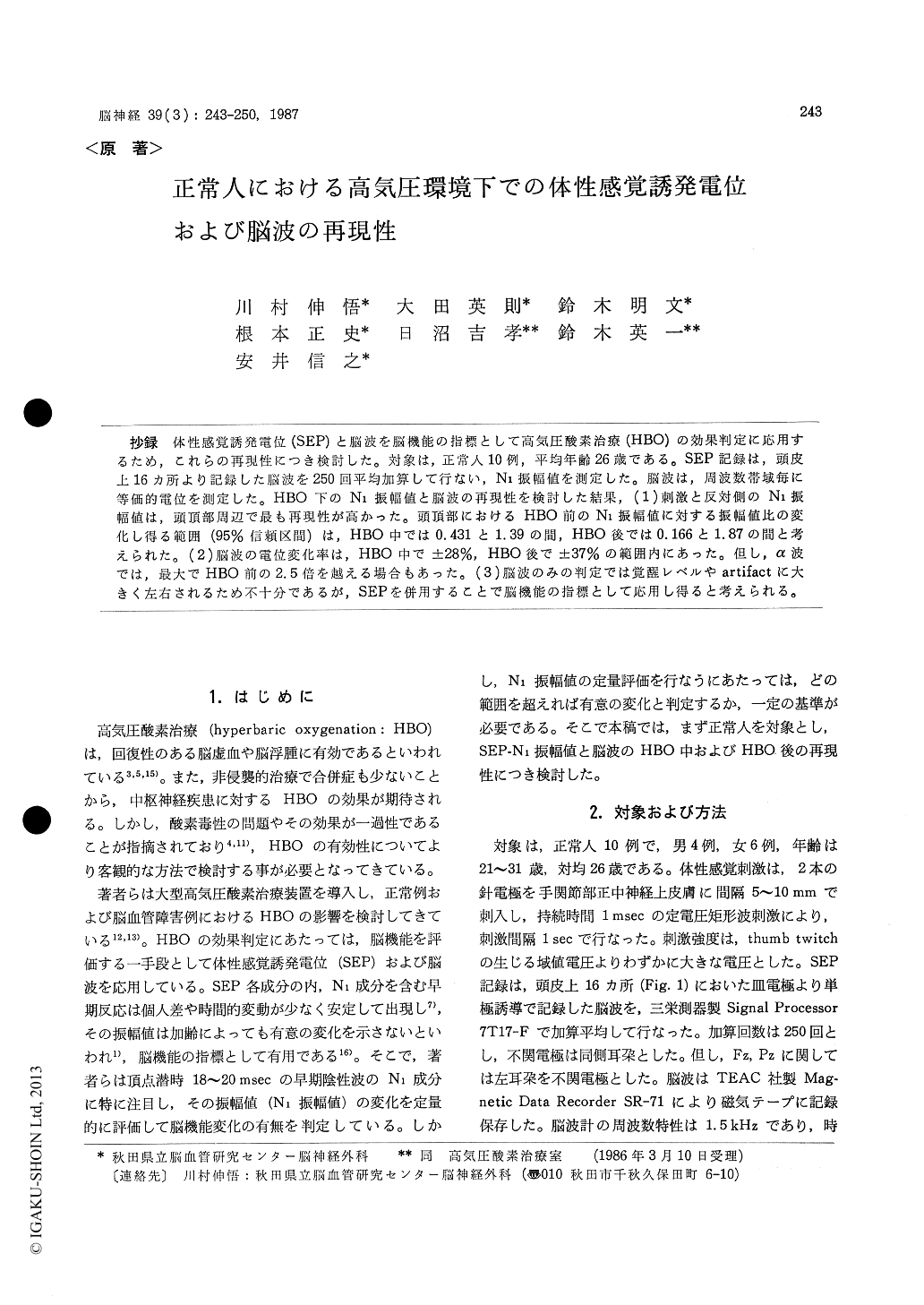
抄録 体性感覚誘発電位(SEP)と脳波を脳機能の指標として高気圧酸素治療(HBO)の効果判定に応用するため,これらの再現性につき検討した。対象は,正常人10例,平均年齢26歳である。SEP記録は,頭皮上16カ所より記録した脳波を250回平均加算して行ない,N1振幅値を測定した。脳波は,周波数帯域毎に等価的電位を測定した。HBO下のN1振幅値と脳波の再現性を検討した結果,(1)刺激と反対側のN1振幅値は,頭頂部周辺で最も再現性が高かった。頭頂部におけるHBO前のN1振幅値に対する振幅値比の変化し得る範囲(95%信頼区間)は,HBO中では0.431と1.39の間,HBO後では0.166と1.87の間と考えられた。(2)脳波の電位変化率は,HBO中で±28%,HBO後で±37%の範囲内にあった。但し,α波では,最大でHBO前の2.5倍を越える場合もあった。(3)脳波のみの判定では覚醒レベルやartifactに大きく左右されるため不十分であるが,SEPを併用することで脳機能の指標として応用し得ると考えられる。
Hyperbaric oxygenation (HBO) was reported tohave a favorable influence on ischemic brain dys-function, and also appeared to be useful in im-proving brain edema. Somatosensory evoked poten-tials (SEP) and EEG would be available as indica-tors of brain function. The purpose of this paper is to study reproducibility of the N1-amplitude of SEP and EEG under HBO.
Materials were 10 normal volunteers, from 21 to 31 (mean age: 26) years old. Two stimulating needle electrodes were inserted into the skin at the wrist and somatosensory stimulations were applied to the median nerve with 1 msec dura-tion square pulses. Stimulation pulses were gene-rated regularly every 1 sec., and stimulus intensity was adjusted just above the thumb twitch thre-shold. Monopolar EEGs were recorded from 16 silver-cup electrodes on the scalp, and SEPs were recorded by averaging 250 responses in those EEGs using computer technique. N1-amplitude was determined by the vertical distance between peaks of the P1 and N1 components. EEGs were analyzed by Fast Fourier Transform and the square root of the averaged power of each frequency band (δ, θ, α1, α2, β) was obtained and evaluated as the equivalent potential. EEGs under the influence of sleeping or artifacts, such as electromyograms, eye lid movements and wandering of the base line, were excluded in evaluating the EEGs. SEPs and EEGs recorded consecutively. The first record was taken before HBO under air breathing at 1ATA, the second during HBO under pure oxygen breath-ing at 2ATA, and the third after HBO under air breathing at 1 ATA once again. N1-amplitude contralateral to the stimulation was most reprodu-cible on the parietal regions (P3 or P4) where the N1-amplitude ratio, the N1-amplitude recorded be-fore HBO compared with that recorded during and after HBO, revealed figures between 0.431 and 1.39 during HBO and between 0.166 and 1.87 after HBO (p<0.05). The percent change of the EEG potential in each frequency band was within ±28% during HBO and ±37% after HBO. But, the maximum percent change of α1 and α2 ex-ceeded these ranges, and the percent change of α1 reached 257% after HBO, compared with its po-tential before HBO. Thus, EEG alone may be insufficient for making judgements on the effects of HBO when the EEG is evaluated using methods involving short time point samplings. Therefore, supplementary information should be provided by evaluating the N1-amplitude of SEP.

Copyright © 1987, Igaku-Shoin Ltd. All rights reserved.


