Japanese
English
- 有料閲覧
- Abstract 文献概要
- 1ページ目 Look Inside
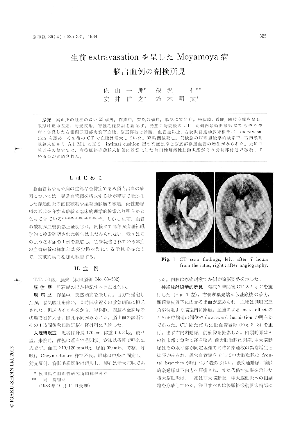
抄録 高血圧の既往のない53歳男。作業中,突然の頭痛,嘔気にて発症。来院時,昏睡,四肢麻痺を呈し,眼球は正中固定。対光反射,脊髄毛様反射を認めず。発症7時間後のCT,両側内頸動脈撮影にてもやもや病に併発した右側頭頭頂部皮質下血腫,脳室穿破と診断。血管撮影上,右後脈絡叢動脈末梢部に,extravasa—tionを認め,その後のCTで血腫は増大していた。53時間後死亡。部検脳の病理し組織学的検索で,右内頸動派終末部からA1 M1に至る,intimal cushion型の高度狭窄と脳底部穿通血管の増生がみられた。更に血腫近傍の検索では,右後脈絡叢動脈末梢部に器質化した陳旧性解離性悩動脈瘤がその分岐部付近で破綻しているのが確認された。
A 53-year-old, non-hypertensive farmer, who had sudden attack of severe headache, was trans-ferred to our clinic. He presented comatous state and tetraparesis without extraocular movements nor reactive pupils to light. CT scan, 7 hours after the ictus showed intracerebral hematoma in the right temporo-parietal region with ventricular extension. The following bilateral carotid angio-grams established the diagnosis of the intracereb-ral hemorrhage due to cerebrovascular moyamoyadisease. In angiograms of the affected side, ir-regular spotty stains spread from the periphei of the right posterior choroidal artery was deli-neated. The repeated CT scan after that indicated increment of the hematoma. Fifty-three hours from the ictus, the patient died and an autopsy study was performed. After the fixation, the coronal brain section was made, and the careful observation of them elucidated the formation of an organized dissecting aneurysm in the angio-graphically extravasated vessel. About seven hund-reds of serial specimen, 4μm in thickness, was then investigated adjacent to the aneurysm. The organized dissecting aneurysm seemed to intiate from the branch of it, where marked fraying and undulation of the fibroelastic intima and internal elastic laminae were observed. The concavity to-ward the true lumen was completely disrupted and communicated to the extravascular space. As a result, the continuous part of it obstructed the lumen of the branch. These findings suggested the newly-developed dissection and it seemed to correspond to the angiographical extravasated points.
Copyright © 1984, Igaku-Shoin Ltd. All rights reserved.