Japanese
English
- 有料閲覧
- Abstract 文献概要
- 1ページ目 Look Inside
I.はじめに
トルコ鞍内およびトルコ鞍近傍は頭蓋底でも最も腫瘍の好発する部位である。そのX線学的診断方法としては,従来頭蓋単純撮影,気脳写,脳血管撮影その他の方法が行われてきた。しかし1972年にコンピューター処理によるX線軸位横断断層撮影装置4)(computerizedtransverse axial tomography)が開発されて以来,本装置は頭蓋内疾患の診断に応用され,現在世界的に同種装置が急速に普及しつつある。
しかしこれらの装置では主として脳横断断層撮影法(Transverse tomography)を中心に開発が行われてきたために,トルコ鞍などの頭蓋底付近の病変の診断には不適当な場合が多かつた。その理由としては頭蓋底骨とほぼ平行にスキヤンニングするため頭蓋底骨とそれに近接する脳病変部との区別が再生画像に明確に表示されないことが多いためであつた。全く同じことが頭頂部のhigh convexity領域にもあてはめられる。これらはいずれも頭蓋骨と装置の物理学的,コンピューター学的諸問題によるものである。
The Craniocervical Computed Tomography wasdeveloped by this author and HITACHI groups inJune, 1976. This author devised the coronal tomo-graphy with the patient in a sitting position.
The scanner completes two adjacent 0.5 or 1.0cm.thick slices in approximately 3 min., 40 sec. Animage reconstruction formed on a 256 × 256 matrixis displayed on monocolor and color Tv monitorsand is recorded on Polaroad Film. The X-ray gene-rator is operated at 120kV and 30mA.
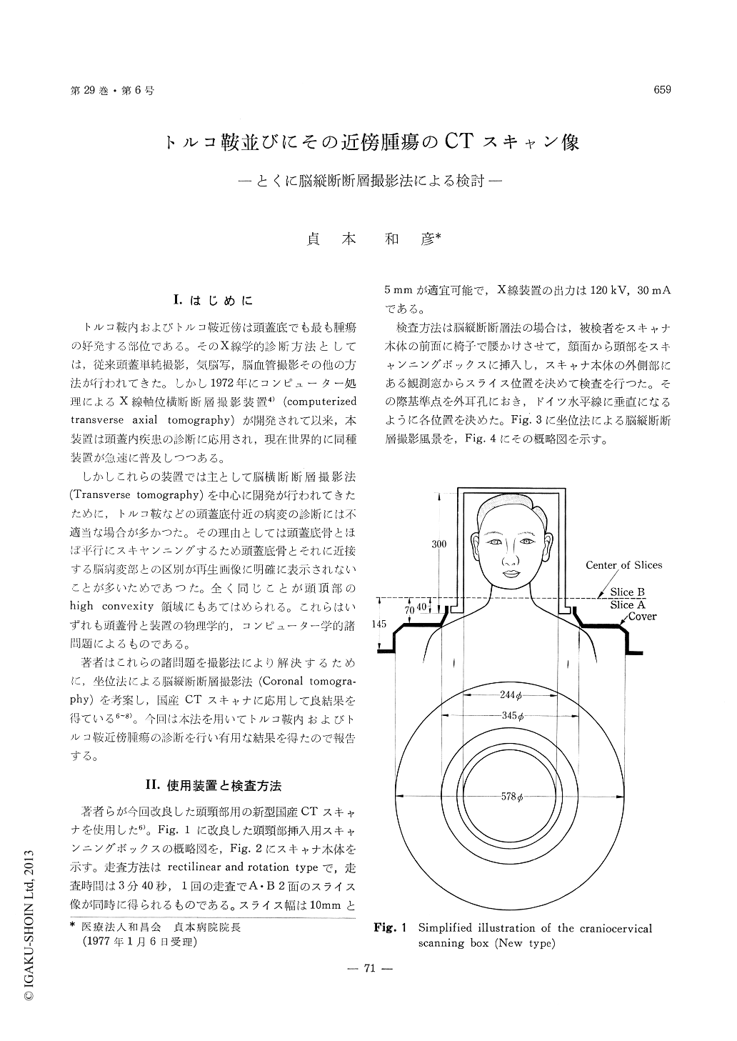
The patient is put in a sitting position on achairin front of the scanner and inserted the head intothe scanning box. The position of the slice ismeasured and decided through the observationwindow which is located at the lateral side of thescanner. The standardpoint and the standardlineare the external auditory meatus and the Reidsbase line.
The sellar and parasellar regions were easilydemonstrated by this coronal tomography. Coronaltomograms in the sellar region were classified intothree types : (1) Anterior type which was sliced atthe anterior part of the sella. (2) Central typewhich was sliced at the central part of the sella.(3) Posterior type which was sliced at the posteriorpart of the sella. These classifications were con-venient for demonstration of each coronal tomogramin the sellar region.
As a result, sellar tumors which were located inthe sella were easily and definitively detected bycoronal tomography, but they could not be detecteddefinitively by transverse tomography. Suprasellartumors were also more easily and definitively de-tected by the coronal tomography than by trans-verse tomography. Furthermore, the relationshipbetween the tumor and the basis (or sella) and be-tween the tumor and the ventricles were also moreclearly differenciated by the coronal tomography.Emphasis was placed on the coronal tomographywhich was most useful for diagnosis of the sellarand parasellar tumors.
Copyright © 1977, Igaku-Shoin Ltd. All rights reserved.