Japanese
English
- 有料閲覧
PPV購入案内
1,320円 (1,200円+税10%) こちらは、911ページから921ページの文献です。購入には医書.jp本会員登録が必要です。
会員登録完了後に改めて上記「購入サイトへ」を選択してください。
または「購入サイトへ」選択後に「購入ログイン」、「新規会員登録」の順に進んでください。
会員登録について詳しくはをご確認ください。
- Abstract 文献概要
- 1ページ目 Look Inside
I.はじめに
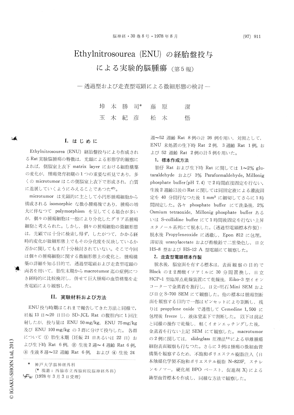
Ethylnitrosourea (ENU)経胎盤投与により作成されるRat実験脳腫瘍の特徴は,光顕による形態学的観察によれば,側脳室上衣下matrix layerにおける細胞構築の変化が,腫瘍発育初期の1つの重要な所見であり,多くのmicrotumorはこの側脳室,上衣下で形成され,白質に進展していくようにみえることであつた47)。
microtumorは光顕的に主として小円形腫瘍細胞から構成されるisomorphicな微小腫瘍塊であり,腫瘍の増大に伴なつてpolymorphismを呈してくる場合が多いが,個々の腫瘍細胞は一般により分化したグリア系腫瘍細胞と考えられた。しかし,個々の腫瘍細胞の微細形態は、光顕では十分に検索し得ず,したがつて,かかる経時的変化が微細形態上でもその分化度を反映しているか否かに関してもまだ十分検討されていない。そこで今回は個々の腫瘍細胞に関する微細形態上の変化と,腫瘍構築の詳細を知る目的で,透過型電顕および走査型電顕の両者を用いて,胎生末期からmacrotumor迄の症例につき経時的に比較検討し,併せて巨大腫瘍の血管構築を走査電顕により観察した。
The fine structure of rat gliomas induced trans-placentally with a single i.p. does of 50 mg/kg of Ethylnitrosourea has been studied by using trans-mission and scanning electron microscope.
The subependymal matrix layers of the fetus which was affected by ENU have showed irregular and rough arrangements with expanded extracellular spaces as compared with that of control rats.
The cells of subependymal layer seemed to form the microtumor. A so-called "microtumor", which was found in a 8 week old, has been composed of small round cells. The fine structures of these cells have showed the characteristics in primitive oligo-dendroglioma.
The characteristics of the fine structure of astro-cytoma cells was identified by both TEM and SEM. The fine structure of subependymal glioma cells was often pleomorphic. These gliomas contained a mixture of primitive oligodendrocytes and epen-dymal cells together with anaplastic glial cells. With increasing size, the glioma has become more pleomorphic with a mixture of neoplastic oligo-dendrocytes, astrocytes and ependymal cells, and ependymoma like cells have showed neither cilia nor junctional complex.
Abnormal vascular structure in the tumor has been reconfirmed by injection replica scanning elec-tron microscope method.
The fine structure of the separated single tumor cell surface was also studied by scanning electron microscope. The differences of the cells surface between that of astrocytoma cell and oligodendro-glioma cells were clearly noticed.
Copyright © 1978, Igaku-Shoin Ltd. All rights reserved.