Japanese
English
- 有料閲覧
PPV購入案内
1,320円 (1,200円+税10%) こちらは、1341ページから1347ページの文献です。購入には医書.jp本会員登録が必要です。
会員登録完了後に改めて上記「購入サイトへ」を選択してください。
または「購入サイトへ」選択後に「購入ログイン」、「新規会員登録」の順に進んでください。
会員登録について詳しくはをご確認ください。
- Abstract 文献概要
- 1ページ目 Look Inside
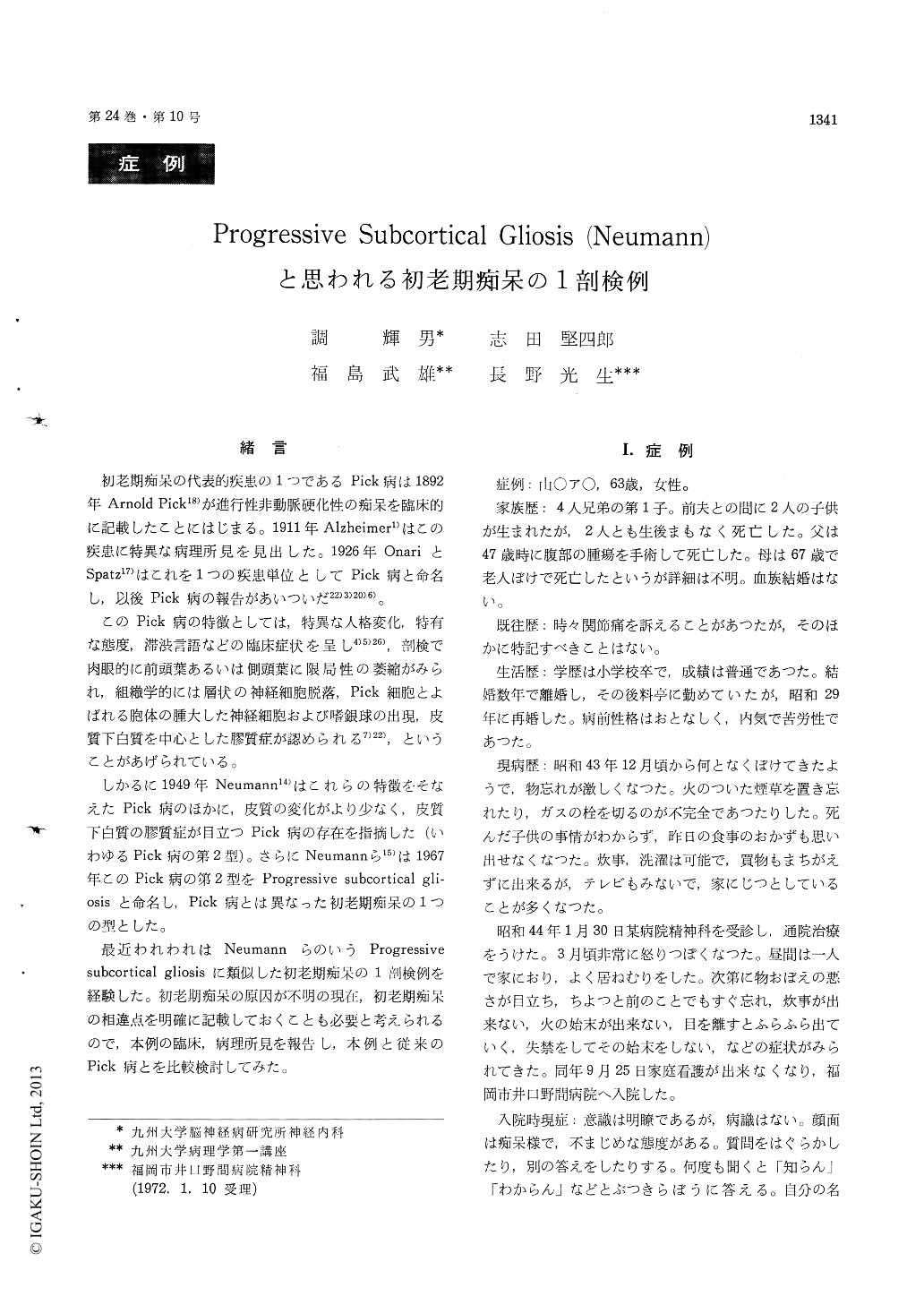
緒言
初老期痴呆の代表的疾患の1つであるPick病は1892年Arnold Pick18)が進行性非動脈硬化性の痴呆を臨床的に記載したことにはじまる。1911年Alzheimer1)はこの疾患に特異な病理所見を見出した。1926年OnariとSpatz17)はこれを1つの疾患単位としてPick病と命名し,以後Pick病の報告があいついだ22)3)20)6)。
このPick病の特徴としては,特異な人格変化,特有な態度,滞渋言語などの臨床症状を呈し4)5)26),剖検で肉眼的に前頭葉あるいは側頭葉に限局性の萎縮がみられ,組織学的には層状の神経細胞脱落,Pick細胞とよばれる胞体の腫大した神経細胞および嗜銀球の出現,皮質下白質を中心とした膠質症が認められる7)22),ということがあげられている。
A 63-year-old woman was admitted to the Noma Psychiatric Hospital, Fukuoka City, in September 1969, for progressive dementia. In December 1968, she had been noticed to become mentally dull. She could not manage the house work and wandered about anytime. At the time of admission, she was alert and unremarkable physically and neurologi-cally except for mild emaciation. Psychiatrically there were disorientation, memory impairment and character change. She was diagnosed as a presenile dementia. After admission, the dementia progress-ed and dirty behavior was seen. In September 1970, logoclonia was detected. Neurological examination at that time revealed rigospasticity in the neck and the extremities. She became bed-ridden there-after and died of bronchopneumonia on April 3, 1971.
At the time of autopsy, the brain weighed 930g. The leptomeninges were thickened and turbid slight-ly. There was mild atrophy in the frontal and temporal lobes on both sides. Moderate dilatation was apparent in the lateral and third ventricles.
With the light microscope, nerve cell loss, spongy change and gliosis in the gray matter were seen in the frontal and temporal lobes. The remaining nerve cells showed simple atrophy, swelling or va-cuolar degeneration. There were mild demyelina-tion and marked gliosis in the white matter in these lobes. Mild or moderate gliosis was seen also in the thalamus. basal ganglia, dentate nucleus, in-ferior olives and the anterior horns in the spinal cord. Senile plaques, neurofibrillary tangles and argyrophilic inclusions were not found in the cent-ral nervous system.
These neuropathological findings were different from those of Alzheimer's disease or Pick's disease and were thought to be akin to the "Progressive Subcortical Gliosis"" reported by Neumann et al. in 1967.
Copyright © 1972, Igaku-Shoin Ltd. All rights reserved.