Japanese
English
- 有料閲覧
PPV購入案内
1,320円 (1,200円+税10%) こちらは、1193ページから1204ページの文献です。購入には医書.jp本会員登録が必要です。
会員登録完了後に改めて上記「購入サイトへ」を選択してください。
または「購入サイトへ」選択後に「購入ログイン」、「新規会員登録」の順に進んでください。
会員登録について詳しくはをご確認ください。
- Abstract 文献概要
- 1ページ目 Look Inside
はじめに
眼球運動については古くから種々の観察が行なわれてきたが,とくに眼振として,小脳機能や前庭機能の表現をみるものとして重要視されてきた。
しかし,例えば逆説睡眠期に現われる速い眼球運動rapid eye movements8)9),入眠期に脳波の変化と平行して出現する遅い動きlangsame Pendeldeviation12),無動無言症の患者で眼だけが不規則に動くことが観察される6),などの現象は,眼球運動が脳の活動水準とかなり密接に関係をもつていることを示唆している。眼球運動の観察は,最終的には中脳の眼球運動核の活動をみていることになるが,さらにさかのぼれば,その核に直接影響を与えている各脳部位の活動をみることになる22)。
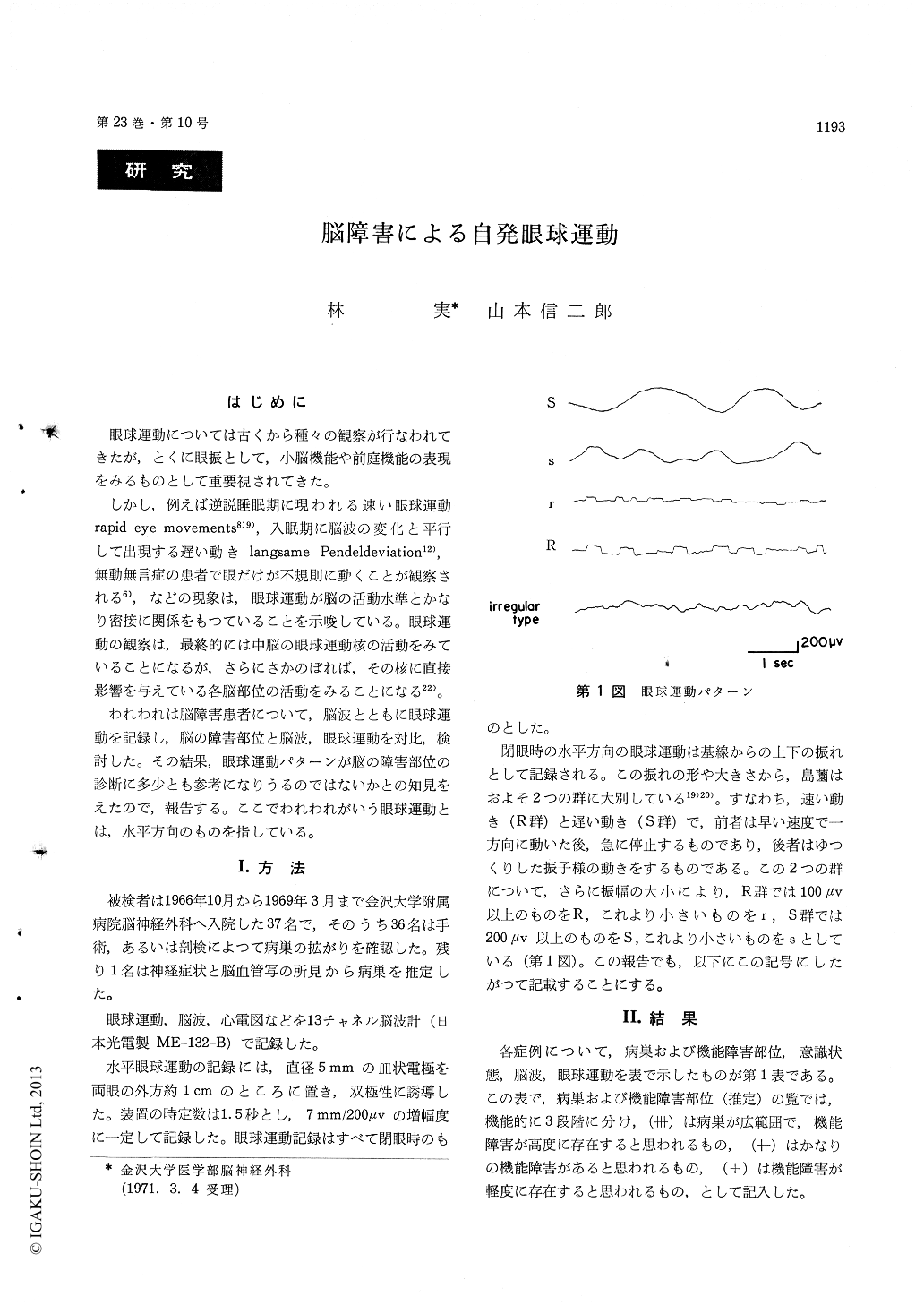
Using 37 brain damaged subjects, we examined electroencephalogram and horizontal eye movements, and correlated these findings with the locations of brain lesions. Horizontal eye movements were classified roughly into 3 main groups, such as slow movements (S-group), rapid movements (R-group) and irregular movements (irregular type).
The eye movements in S-group appeared generally when the lesion was located in the telencephalon and diencephalon.
Those in R-group tended to appear in the brain-stem, especially in the pons lesion.
Those in irregular type occured in the lession of medulla oblongata.
Eye movement disappeared when the lesion was located in the mesencephalon.
Copyright © 1971, Igaku-Shoin Ltd. All rights reserved.