Japanese
English
- 有料閲覧
PPV購入案内
1,320円 (1,200円+税10%) こちらは、19ページから23ページの文献です。購入には医書.jp本会員登録が必要です。
会員登録完了後に改めて上記「購入サイトへ」を選択してください。
または「購入サイトへ」選択後に「購入ログイン」、「新規会員登録」の順に進んでください。
会員登録について詳しくはをご確認ください。
- Abstract 文献概要
- 1ページ目 Look Inside
序言
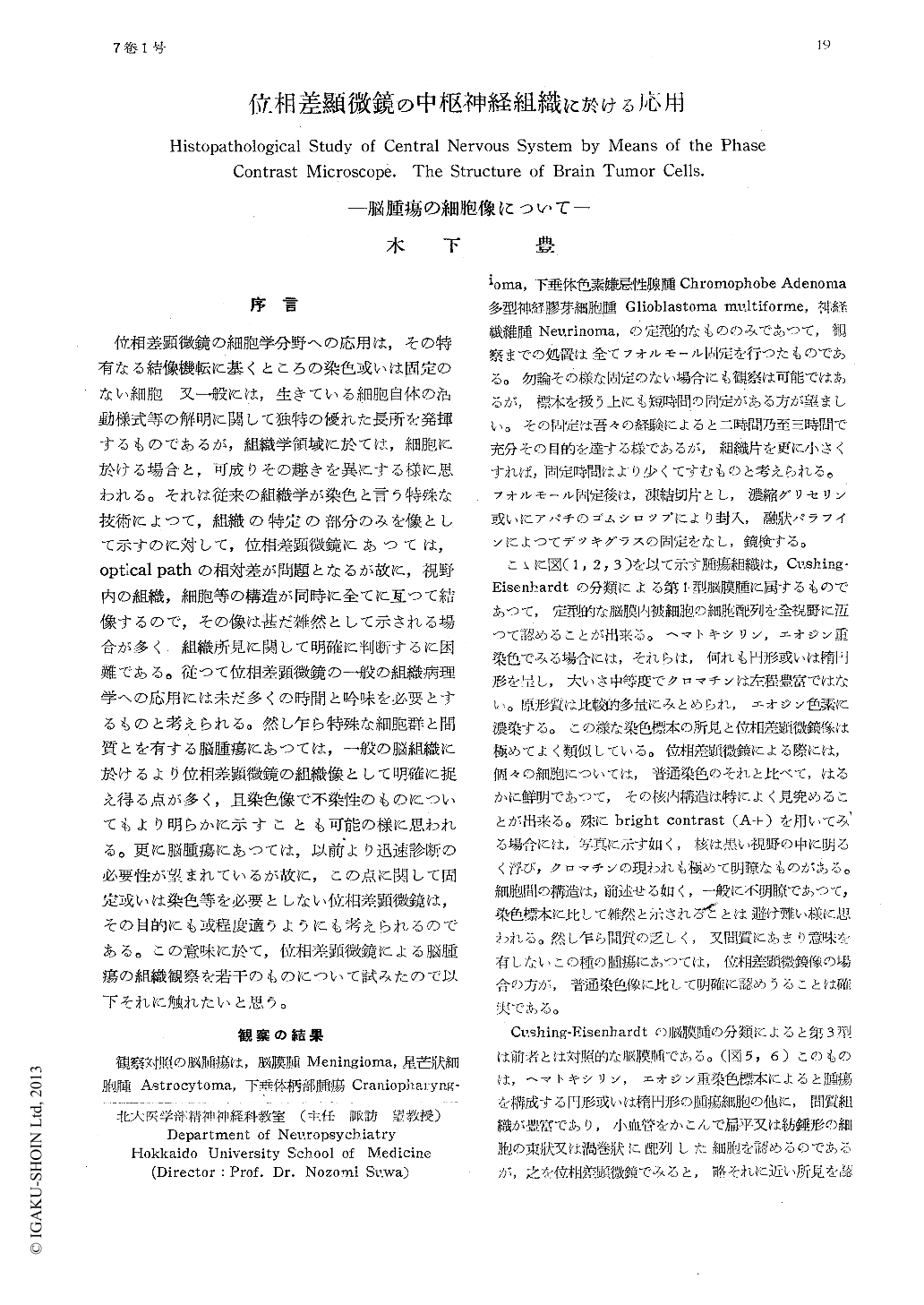
位相差顕微鏡の細胞学分野への応用は,その特有なる結像機転に基くところの染色或いは固定のない細胞 又一般には,生きている細胞自体の活動様式等の解明に関して独特の優れた長所を発揮するものであるが,組織学領域に於ては,細胞に於ける場合と,可成りその趣きを異にする様に思われる。それは従来の組織学が染色と言う特殊な技術によつて,組織の特定の部分のみを像として示すのに対して,位相差顕微鏡にあつては,optical pathの相対差が問題となるが故に,視野内の組織,細胞等の講造が同時に全てに亙つて結像するので,その像は甚だ雑然として示される場合が多く 組織所見に関して明確に判断するに困難である。従つて位相差顕微鏡の一般の組織病理学への応用には未だ多くの時間と吟味を必要とするものと考えられる。然し乍ら特殊な細胞群と間質とを有する脳腫瘍にあつては,一般の脳組織に於けるより位相差顕微鏡の組織像として明確に捉え得る点が多く,且染色像で不染性のものについてもより明らかに示すことも可能の様に思われる。更に脳腫瘍にあつては,以前より迅速診断の必要性が望まれているが故に,この点に関して固定或いは染色等を必要としない位相差顕微鏡は,その目的にも或程度適うようにも考えられるのである。この意味に於て,位相差顕微鏡による脳腫瘍の組織観察を若干のものについて試みたので以下それに触れたいと思う。
The cellular structure of various kinds of brain tumors was investigated by means of the phase contrast microscope.
In the case of meningioma, craniopharyn-giorna, chromophobe adenoma and astrocy-toma, it was possible to determine their histopathological diagnosis by verifying the charateritic arrangement, structure and type of their tumor cells. It could be concluded, therefore, that it is possible to diagnose the several kind of brain tumors histpathologica-lly with certain accuracy without any stain, which would be significant from the clinical point of view.
Copyright © 1955, Igaku-Shoin Ltd. All rights reserved.