Japanese
English
- 有料閲覧
PPV購入案内
1,320円 (1,200円+税10%) こちらは、305ページから308ページの文献です。購入には医書.jp本会員登録が必要です。
会員登録完了後に改めて上記「購入サイトへ」を選択してください。
または「購入サイトへ」選択後に「購入ログイン」、「新規会員登録」の順に進んでください。
会員登録について詳しくはをご確認ください。
- Abstract 文献概要
- 1ページ目 Look Inside
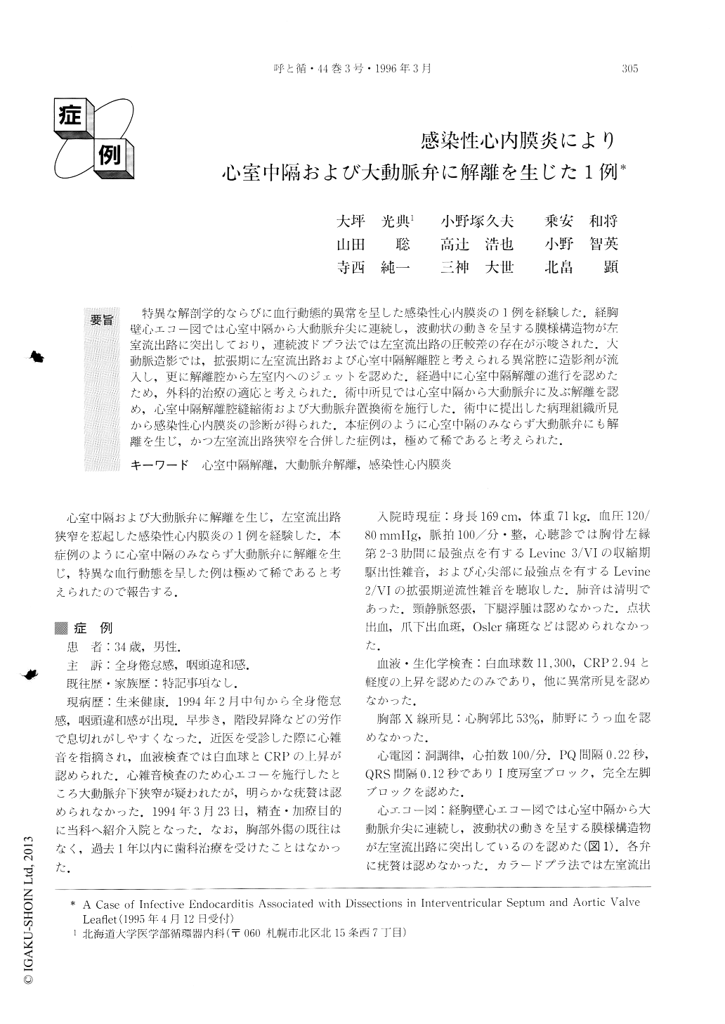
特異な解剖学的ならびに血行動態的異常を呈した感染性心内膜炎の1例を経験した.経胸壁心エコー図では心室中隔から大動脈弁尖に連続し,波動状の動きを呈する膜様構造物が左室流出路に突出しており,連続波ドプラ法では左室流出路の圧較差の存在が示唆された.大動脈造影では,拡張期に左室流出路および心室中隔解離腔と考えられる異常腔に造影剤が流入し,更に解離腔から左室内へのジェットを認めた.経過中に心室中隔解離の進行を認めたため,外科的治療の適応と考えられた.術中所見では心室中隔から大動脈弁に及ぶ解離を認め,心室中隔解離腔縫縮術および大動脈弁置換術を施行した.術中に提出した病理組織所見から感染性心内膜炎の診断が得られた.本症例のように心室中隔のみならず大動脈弁にも解離を生じ,かつ左室流出路狭窄を合併した症例は,極めて稀であると考えられた.
We report a very rare case of infective endocarditis with dissections both in the interventricular septum and in an aortic valve leaflet.
A swinging membranous structure was detected in the left ventricular outflow tract by transthoracic and transesophageal echocardiography, and it was thought to he caused by a dissection of the interventricular septum. Doppler technique revealed a significant systolic pressure gradient across the abnormal structure with an aortic regurgitant jet. Aortography showed aortic regurgitation, and the interventricular dissected zone was enhanced at the diastolic phase. There was a jet of contrast from the dissected zone to the left ventricule.
We detected the extension of the dissected area by echocardiography after the hospitalization, and the patient was operated on. Dissections both in the inter-ventricular septum and the aortic valve leaflet were observed at the operation and the pathological exami-nation was compatible with healed infective endocar-ditis.
Copyright © 1996, Igaku-Shoin Ltd. All rights reserved.