Japanese
English
- 有料閲覧
PPV購入案内
1,320円 (1,200円+税10%) こちらは、367ページから373ページの文献です。購入には医書.jp本会員登録が必要です。
会員登録完了後に改めて上記「購入サイトへ」を選択してください。
または「購入サイトへ」選択後に「購入ログイン」、「新規会員登録」の順に進んでください。
会員登録について詳しくはをご確認ください。
- Abstract 文献概要
- 1ページ目 Look Inside
- 参考文献 Reference
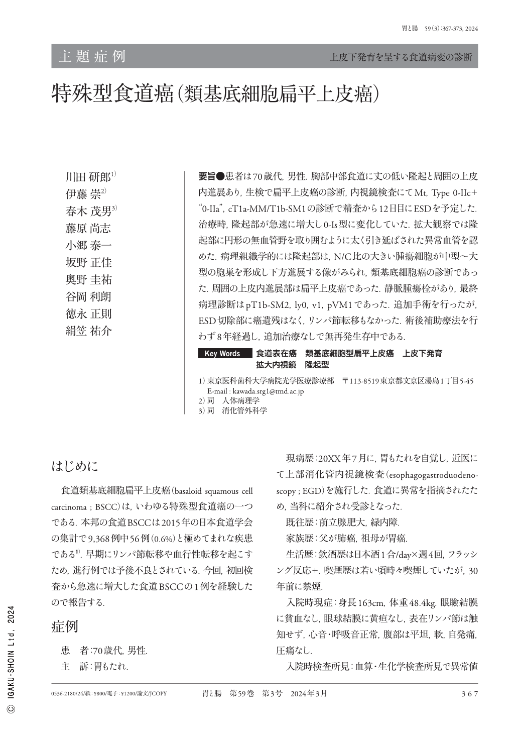
要旨●患者は70歳代,男性.胸部中部食道に丈の低い隆起と周囲の上皮内進展あり,生検で扁平上皮癌の診断,内視鏡検査にてMt,Type 0-IIc+“0-IIa”,cT1a-MM/T1b-SM1の診断で精査から12日目にESDを予定した.治療時,隆起部が急速に増大し0-Is型に変化していた.拡大観察では隆起部に円形の無血管野を取り囲むように太く引き延ばされた異常血管を認めた.病理組織学的には隆起部は,N/C比の大きい腫瘍細胞が中型〜大型の胞巣を形成し下方進展する像がみられ,類基底細胞癌の診断であった.周囲の上皮内進展部は扁平上皮癌であった.静脈腫瘍栓があり,最終病理診断はpT1b-SM2,ly0,v1,pVM1であった.追加手術を行ったが,ESD切除部に癌遺残はなく,リンパ節転移もなかった.術後補助療法を行わず8年経過し,追加治療なしで無再発生存中である.
A 70-year-old male underwent esophagogastroduodenoscopy. A flat elevated lesion surrounded by intraepithelial invasion was observed in the middle esophagus. The biopsy specimen led to the diagnosis of squamous cell carcinoma. The depth of tumor was diagnosed as T1a-MM or T1b-SM1 ; thus, ESD was performed. Evaluation 12 days after initial observation revealed that the elevated lesion grew up to the protruded lesion.Magnifying endoscopy revealed avascular areas with unusual surrounding vessels. The lesion was endoscopically resected, and the tumor was histologically diagnosed as basaloid(squamous)carcinoma, SM2(1700μm), ly0, v1, HM0, VM+. Additional subtotal esophagectomy with lymph node resection was performed, and no residual lesion or lymph node metastasis was found. He received no adjuvant treatment and has survived for 8 years without recurrence.
Copyright © 2024, Igaku-Shoin Ltd. All rights reserved.