Japanese
English
- 有料閲覧
PPV購入案内
1,320円 (1,200円+税10%) こちらは、1120ページから1127ページの文献です。購入には医書.jp本会員登録が必要です。
会員登録完了後に改めて上記「購入サイトへ」を選択してください。
または「購入サイトへ」選択後に「購入ログイン」、「新規会員登録」の順に進んでください。
会員登録について詳しくはをご確認ください。
- Abstract 文献概要
- 1ページ目 Look Inside
- 参考文献 Reference
- サイト内被引用 Cited by
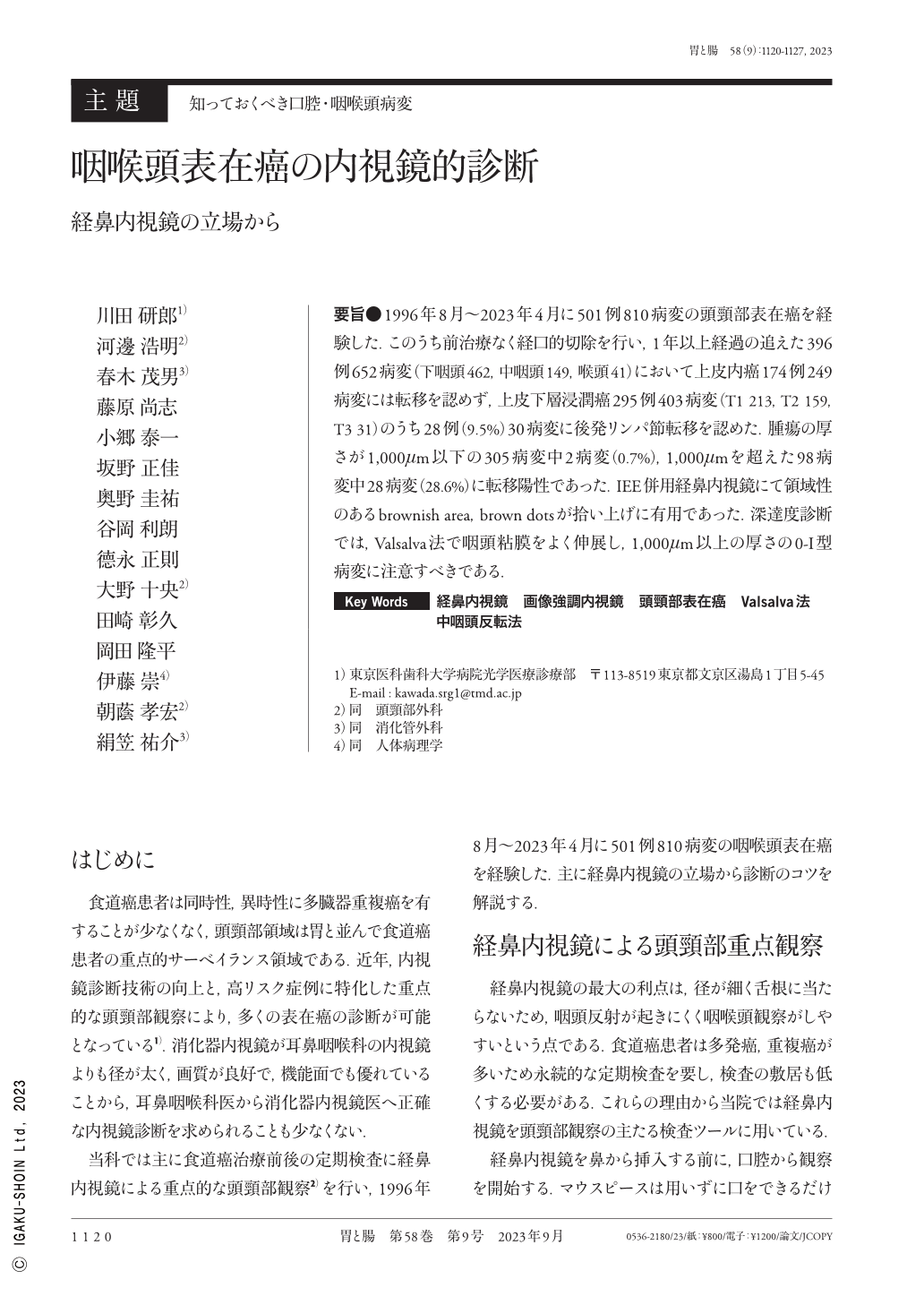
要旨●1996年8月〜2023年4月に501例810病変の頭頸部表在癌を経験した.このうち前治療なく経口的切除を行い,1年以上経過の追えた396例652病変(下咽頭462,中咽頭149,喉頭41)において上皮内癌174例249病変には転移を認めず,上皮下層浸潤癌295例403病変(T1 213,T2 159,T3 31)のうち28例(9.5%)30病変に後発リンパ節転移を認めた.腫瘍の厚さが1,000μm以下の305病変中2病変(0.7%),1,000μmを超えた98病変中28病変(28.6%)に転移陽性であった.IEE併用経鼻内視鏡にて領域性のあるbrownish area,brown dotsが拾い上げに有用であった.深達度診断では,Valsalva法で咽頭粘膜をよく伸展し,1,000μm以上の厚さの0-I型病変に注意すべきである.
We experienced 501 cases of superficial carcinoma of the head and neck with 810 lesions from August 1996 to April 2023. Of these, 652 lesions in 396 cases were with endoscopic resection and without prior treatment and follow-up of >1 year(462 lesions of hypopharynx, 149 lesions of oropharynx, and 41 lesions of larynx), 249 lesions in 174 cases with intraepithelial carcinoma showed no metastasis, and 30 lesions in 28 cases(9.5%)out of 403 lesions(T1:213, T2:159, T3:31)in 295 cases with subepithelial invasive carcinoma showed metastasis. Metastasis was positive in 2 of 305(0.7%)lesions with tumor thickness <1,000μm and 28 of 98(28.6%)lesions with tumor thickness >1,000μm. Nasal endoscopy with image-enhanced endoscopy was useful in identifying brownish areas and brown dots. The pharyngeal mucosa should be well stretched using the Valsalva method for depth diagnosis, and type 0−I lesions with a thickness of >1,000μm should be noted.
Copyright © 2023, Igaku-Shoin Ltd. All rights reserved.