Japanese
English
- 有料閲覧
PPV購入案内
1,320円 (1,200円+税10%) こちらは、1685ページから1693ページの文献です。購入には医書.jp本会員登録が必要です。
会員登録完了後に改めて上記「購入サイトへ」を選択してください。
または「購入サイトへ」選択後に「購入ログイン」、「新規会員登録」の順に進んでください。
会員登録について詳しくはをご確認ください。
- Abstract 文献概要
- 1ページ目 Look Inside
- 参考文献 Reference
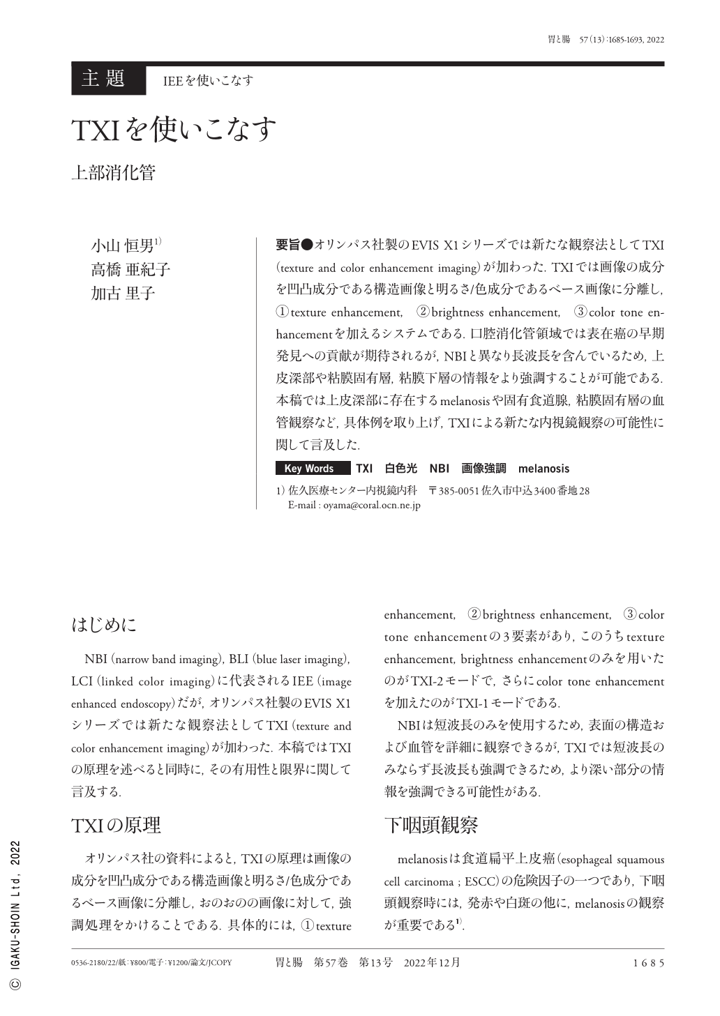
要旨●オリンパス社製のEVIS X1シリーズでは新たな観察法としてTXI(texture and color enhancement imaging)が加わった.TXIでは画像の成分を凹凸成分である構造画像と明るさ/?成分であるベース画像に分離し,①texture enhancement,②brightness enhancement,③color tone enhancementを加えるシステムである.口腔消化管領域では表在癌の早期発見への貢献が期待されるが,NBIと異なり長波長を含んでいるため,上皮深部や粘膜固有層,粘膜下層の情報をより強調することが可能である.本稿では上皮深部に存在するmelanosisや固有食道腺,粘膜固有層の血管観察など,具体例を取り上げ,TXIによる新たな内視鏡観察の可能性に関して言及した.
TXI(texture and color enhancement imaging)provides better visibility of potential and extant lesions by enhancing the texture, brightness, and color to define subtle tissue differences more clearly. This tool is expected to contribute to the early detection of superficial cancer in the oral cavity and gastrointestinal tract. Moreover, unlike narrow band imaging, it contains long wavelengths ; therefore, information on the deep epithelium, lamina propria, and submucosa can be emphasized. We have mentioned the possibility of new endoscopic observations using TXI, considering specific examples, such as the assessment of melanosis existing in the deep epithelium, proper esophageal gland, and blood vessel observation of the lamina propria.
Copyright © 2022, Igaku-Shoin Ltd. All rights reserved.