Japanese
English
- 有料閲覧
PPV購入案内
1,320円 (1,200円+税10%) こちらは、1617ページから1625ページの文献です。購入には医書.jp本会員登録が必要です。
会員登録完了後に改めて上記「購入サイトへ」を選択してください。
または「購入サイトへ」選択後に「購入ログイン」、「新規会員登録」の順に進んでください。
会員登録について詳しくはをご確認ください。
- Abstract 文献概要
- 1ページ目 Look Inside
- 参考文献 Reference
- サイト内被引用 Cited by
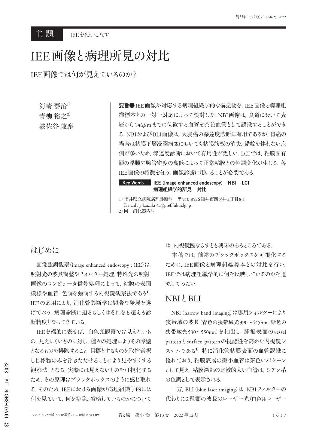
要旨●IEE画像が対応する病理組織学的な構造物を,IEE画像と病理組織標本との一対一対応によって検討した.NBI画像は,食道において表層から146μmまでに位置する血管を茶色血管として認識することができる.NBIおよびBLI画像は,大腸癌の深達度診断に有用であるが,胃癌の場合は粘膜下層浸潤病変においても粘膜筋板の消失,錯綜を伴わない症例が多いため,深達度診断において有用性が乏しい.LCIでは,粘膜固有層の浮腫や腺管密度の高低によって正常粘膜との色調変化が生じる.各IEE画像の特徴を知り,画像診断に用いることが必要である.
The relationship between IEE(image enhanced endoscopy)images and histopathological specimens was investigated by one-by-one correspondence. NBI(narrow band imaging)images can recognize blood vessels located up to 146μm from the esophageal surface as brown blood vessels. NBI and blue laser imaging images are used for diagnosing the depth of colorectal cancer invasion. In gastric cancer, many patients had submucosal infiltration lesions that do not involve the loss of or entangled muscularis mucosae ; thus, IEE imaging is only slightly used in diagnosing the depth of invasion. In linked color imaging, lamina propria edema and low-density ducts can change the colors of the mucosa. It is necessary to know the characteristics of each IEE image and use them for diagnosis.
Copyright © 2022, Igaku-Shoin Ltd. All rights reserved.