Japanese
English
- 有料閲覧
- Abstract 文献概要
- 1ページ目 Look Inside
- 参考文献 Reference
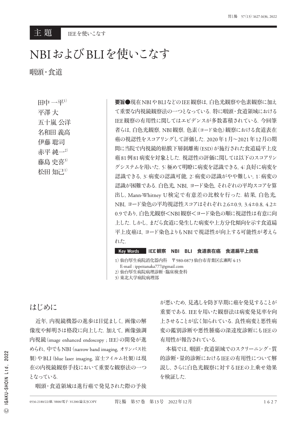
要旨●現在NBIやBLIなどのIEE観察は,白色光観察や色素観察に加えて重要な内視鏡観察法の一つとなっている.特に咽頭・食道領域におけるIEE観察の有用性に関してはエビデンスが多数蓄積されている.今回筆者らは,白色光観察,NBI観察,色素(ヨード染色)観察における食道表在癌の視認性をスコアリングして評価した.2020年1月〜2021年12月の期間に当院で内視鏡的粘膜下層剝離術(ESD)が施行された食道扁平上皮癌81例81病変を対象とした.視認性の評価に関しては以下のスコアリングシステムを用いた.5:極めて明瞭に病変を認識できる,4:良好に病変を認識できる,3:病変の認識可能,2:病変の認識がやや難しい,1:病変の認識が困難である.白色光,NBI,ヨード染色,それぞれの平均スコアを算出し,Mann-Whitney U検定で有意差の比較を行った.結果,白色光,NBI,ヨード染色の平均視認性スコアはそれぞれ2.6±0.9,3.4±0.8,4.2±0.9であり,白色光観察<NBI観察<ヨード染色の順に視認性は有意に向上した.しかし,まだら食道に発生した病変や上方分化傾向を示す食道扁平上皮癌は,ヨード染色よりもNBIで視認性が向上する可能性が考えられた.
One of the most important endoscopic observation methods is IEE(image enhanced endoscopy), which includes NBI(narrow band imaging)and blue laser imaging. Many previous studies have demonstrated the utility of IEE, beginning with Muto et al. paper, which showed that NBI improves the detection rate of superficial cancers of the pharynx and esophagus. Based on the previous studies, we explain the utility of IEE observation for screening, differential diagnosis, and diagnosis of invasion depth of lesions in the pharyngeal and esophageal areas in this paper. Furthermore, we scored and evaluated the visibility of superficial esophageal carcinoma using WLI(white light imaging), NBI, and iodine staining. When iodine staining was used instead of WLI or NBI, the visibility of superficial esophageal carcinoma was significantly improved. However, lesions arising in the esophagus showing multiple iodine voiding lesions after iodine staining, including carcinoma with the superficial layer containing glycogen granules, may be more visible with NBI than with iodine, emphasizing the importance of a comprehensive diagnosis using multiple modalities.

Copyright © 2022, Igaku-Shoin Ltd. All rights reserved.


