Japanese
English
- 有料閲覧
PPV購入案内
1,320円 (1,200円+税10%) こちらは、909ページから917ページの文献です。購入には医書.jp本会員登録が必要です。
会員登録完了後に改めて上記「購入サイトへ」を選択してください。
または「購入サイトへ」選択後に「購入ログイン」、「新規会員登録」の順に進んでください。
会員登録について詳しくはをご確認ください。
- Abstract 文献概要
- 1ページ目 Look Inside
- 参考文献 Reference
- サイト内被引用 Cited by
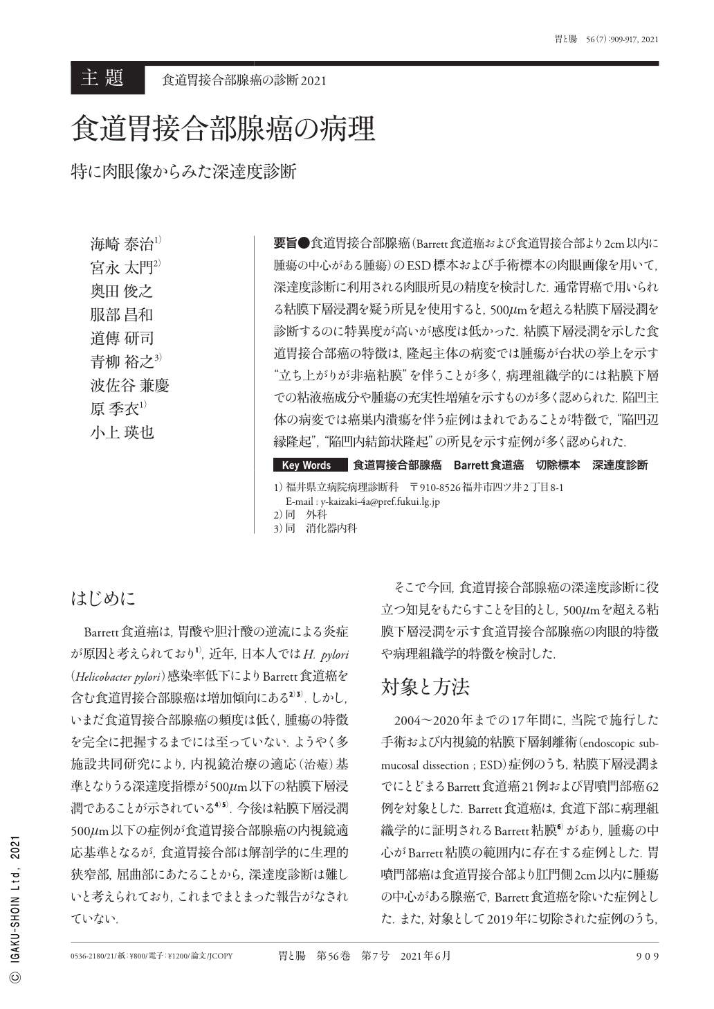
要旨●食道胃接合部腺癌(Barrett食道癌および食道胃接合部より2cm以内に腫瘍の中心がある腫瘍)のESD標本および手術標本の肉眼画像を用いて,深達度診断に利用される肉眼所見の精度を検討した.通常胃癌で用いられる粘膜下層浸潤を疑う所見を使用すると,500μmを超える粘膜下層浸潤を診断するのに特異度が高いが感度は低かった.粘膜下層浸潤を示した食道胃接合部癌の特徴は,隆起主体の病変では腫瘍が台状の挙上を示す“立ち上がりが非癌粘膜”を伴うことが多く,病理組織学的には粘膜下層での粘液癌成分や腫瘍の充実性増殖を示すものが多く認められた.陥凹主体の病変では癌巣内潰瘍を伴う症例はまれであることが特徴で,“陥凹辺縁隆起”,“陥凹内結節状隆起”の所見を示す症例が多く認められた.
We examined the accuracy of macroscopic findings to diagnose the depth of invasion in esophagogastric junction adenocarcinoma using photographs of resected specimens obtained from endoscopic submucosal dissection or surgery. Based on the findings suggesting submucosal invasion in gastric cancer, the specificity of diagnosing submucosal invasion deeper than 500μm was high, but the sensitivity was low. A protruded lesion with a “table-like” appearance is characteristic of esophagogastric junction carcinoma with submucosal invasion. Histologically, lesions showed mucinous carcinoma or solid tumor growth. In addition, the depressed lesions were rarely accompanied by ulcerations. A “recessed marginal ridge” and a “nodular lesion in the depression” were additional findings of esophagogastric junction adenocarcinoma.
Copyright © 2021, Igaku-Shoin Ltd. All rights reserved.