Japanese
English
- 有料閲覧
PPV購入案内
1,320円 (1,200円+税10%) こちらは、1243ページから1252ページの文献です。購入には医書.jp本会員登録が必要です。
会員登録完了後に改めて上記「購入サイトへ」を選択してください。
または「購入サイトへ」選択後に「購入ログイン」、「新規会員登録」の順に進んでください。
会員登録について詳しくはをご確認ください。
- Abstract 文献概要
- 1ページ目 Look Inside
- 参考文献 Reference
- サイト内被引用 Cited by
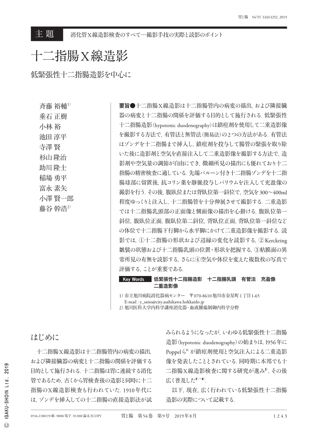
要旨●十二指腸X線造影は十二指腸管内の病変の描出,および隣接臓器の病変と十二指腸の関係を評価する目的として施行される.低緊張性十二指腸造影(hypotonic duodenography)は鎮痙剤を使用して二重造影像を撮影する方法で,有管法と無管法(簡易法)の2つの方法がある.有管法はゾンデを十二指腸まで挿入し,鎮痙剤を投与して腸管の緊張を取り除いた後に造影剤と空気を直接注入して二重造影像を撮影する方法で,造影剤や空気量の調節が自由にでき,微細所見の描出にも優れており十二指腸の精密検査に適している.先端バルーン付き十二指腸ゾンデを十二指腸球部に留置後,抗コリン薬を静脈投与しバリウムを注入して充盈像の撮影を行う.その後,腹臥位または背臥位第一斜位で,空気を300〜400ml程度ゆっくりと注入し,十二指腸管を十分伸展させて撮影する.二重造影では十二指腸乳頭部の正面像と側面像の描出を心掛ける.腹臥位第一斜位,腹臥位正面,腹臥位第二斜位,背臥位正面,背臥位第一斜位などの体位で十二指腸下行脚から水平脚にかけて二重造影像を撮影する.読影では,①十二指腸の形状および辺縁の変化を読影する,②Kerckring皺襞の状態および十二指腸乳頭の位置・形状を把握する,③粘膜面の異常所見の有無を読影する,さらに④空気や体位を変えた複数枚の写真で評価する,ことが重要である.
Radiographic examination of the duodenum is performed to delineate intraduodenal lesions and to evaluate the extent of the disease and involvement of adjacent organs. Hypotonic duodenography is a radiographic procedure that obtains double-contrast images under hypotonic conditions by administering an antispasmodic drug. This technique has two types:tube-assisted and tubeless. For detailed examinations, the tube-assisted method is suitable because minute findings can be obtained by adjusting the amount of barium and air. Hypotonic duodenography is performed by intubation of the balloon tube into the duodenum, followed by inflation of the balloon to fix the position of the tube in the duodenal bulb. An antispasmodic drug is then administered to the patient, followed by slow injection of barium through the tube to obtain barium-filled images. Next, in the prone or supine 1st oblique position, 300-400mL of air is injected until the duodenum lumen is adequately extended and double-contrast images are obtained. The purpose of the double-contrast study should be to visualize the frontal and lateral images of the duodenal papilla. In addition, double-contrast images can be obtained in the following positions:prone, prone 1st and 2nd oblique, supine, and supine 1st and 2nd oblique. Interpretation of the results depends on:(1)duodenal shape and changes of the duodenal border,(2)abnormality of the Kerckring folds and location and shape of the duodenal papilla,(3)abnormality of the duodenal mucosa(conversing mucosal folds, polypoid lesions, and erosions or ulcers), and(4)diagnosis determined after evaluating the multiple images in various positions and amounts of air.
Copyright © 2019, Igaku-Shoin Ltd. All rights reserved.