Japanese
English
- 有料閲覧
PPV購入案内
1,320円 (1,200円+税10%) こちらは、1384ページから1392ページの文献です。購入には医書.jp本会員登録が必要です。
会員登録完了後に改めて上記「購入サイトへ」を選択してください。
または「購入サイトへ」選択後に「購入ログイン」、「新規会員登録」の順に進んでください。
会員登録について詳しくはをご確認ください。
- Abstract 文献概要
- 1ページ目 Look Inside
- 参考文献 Reference
- サイト内被引用 Cited by
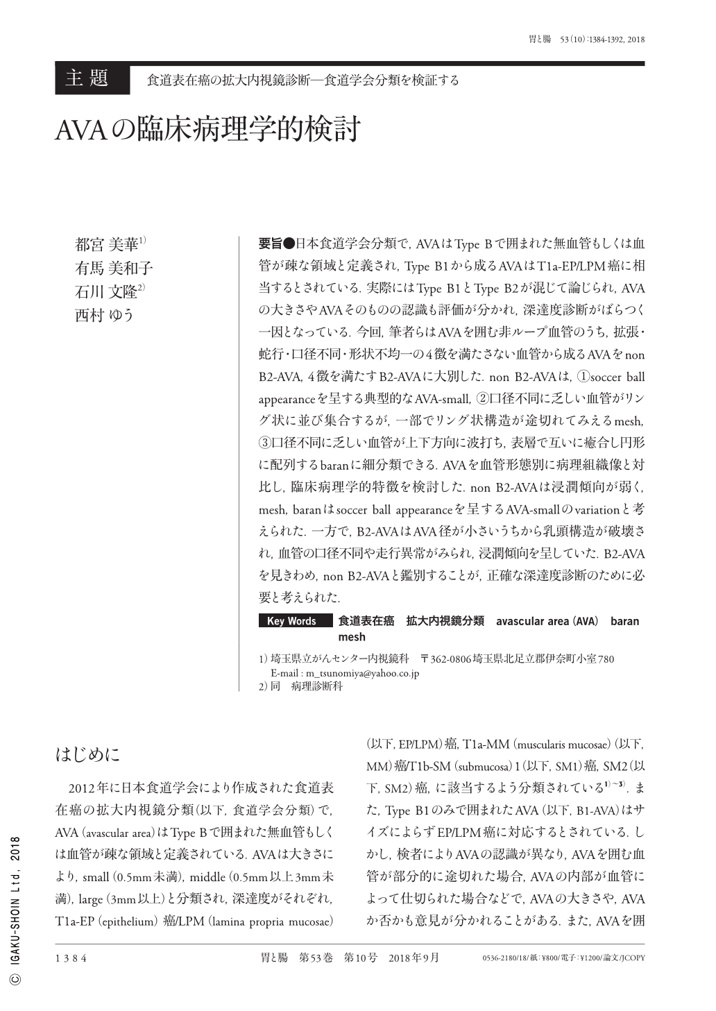
要旨●日本食道学会分類で,AVAはType Bで囲まれた無血管もしくは血管が疎な領域と定義され,Type B1から成るAVAはT1a-EP/LPM癌に相当するとされている.実際にはType B1とType B2が混じて論じられ,AVAの大きさやAVAそのものの認識も評価が分かれ,深達度診断がばらつく一因となっている.今回,筆者らはAVAを囲む非ループ血管のうち,拡張・蛇行・口径不同・形状不均一の4徴を満たさない血管から成るAVAをnon B2-AVA,4徴を満たすB2-AVAに大別した.non B2-AVAは,①soccer ball appearanceを呈する典型的なAVA-small,②口径不同に乏しい血管がリング状に並び集合するが,一部でリング状構造が途切れてみえるmesh,③口径不同に乏しい血管が上下方向に波打ち,表層で互いに癒合し円形に配列するbaranに細分類できる.AVAを血管形態別に病理組織像と対比し,臨床病理学的特徴を検討した.non B2-AVAは浸潤傾向が弱く,mesh,baranはsoccer ball appearanceを呈するAVA-smallのvariationと考えられた.一方で,B2-AVAはAVA径が小さいうちから乳頭構造が破壊され,血管の口径不同や走行異常がみられ,浸潤傾向を呈していた.B2-AVAを見きわめ,non B2-AVAと鑑別することが,正確な深達度診断のために必要と考えられた.
The Japan Esophageal Society uses magnifying endoscopic classification to define AVA as a low or no vascularity area surrounded by type B vessels. Any type of AVA(small, medium, or large)surrounded by type B1 vessels is suggestive of T1a-EP or T1a-LPM squamous cell carcinoma. However, in reality, type B1 and B2 vessels surrounding the AVA are often confused. Recognition of the size of the AVA and the AVA itself varies from person to person. This chaos leads to variance in diagnosis of invasion depth. Here, we classified non-loop vessels surrounding the AVA into non B2-AVA and B2-AVA according to the presence of a tetrad of morphological factors of the vessels:dilation, weaving, irregular caliber, and different shape. Non B2-AVAs are subclassified into typical AVAs:small, of a soccer ball appearance ; mesh, wherein there are ring-like vessels around AVAs that accumulate and part of the ring structure is broken ; and baran, wherein baran-like vessels around the AVA wave vertically and fuse on the surface. We subsequently investigated the pathology and studied the clinicopathological features of the classified AVAs. Non B2-AVA is less invasive. Mesh and baran AVAs are thought to be variations of the AVA-small with soccer ball appearance. Conversely, B2-AVAs have invasive characteristics:the subepithelial papillary structure is broken and the caliber and direction of the vessels is irregular. Extracting B2-AVAs and differentiating them from non B2-AVAs is essential for accurately diagnosing the invasion depth of superficial esophageal cancer.
Copyright © 2018, Igaku-Shoin Ltd. All rights reserved.