Japanese
English
- 有料閲覧
- Abstract 文献概要
- 1ページ目 Look Inside
- 参考文献 Reference
- サイト内被引用 Cited by
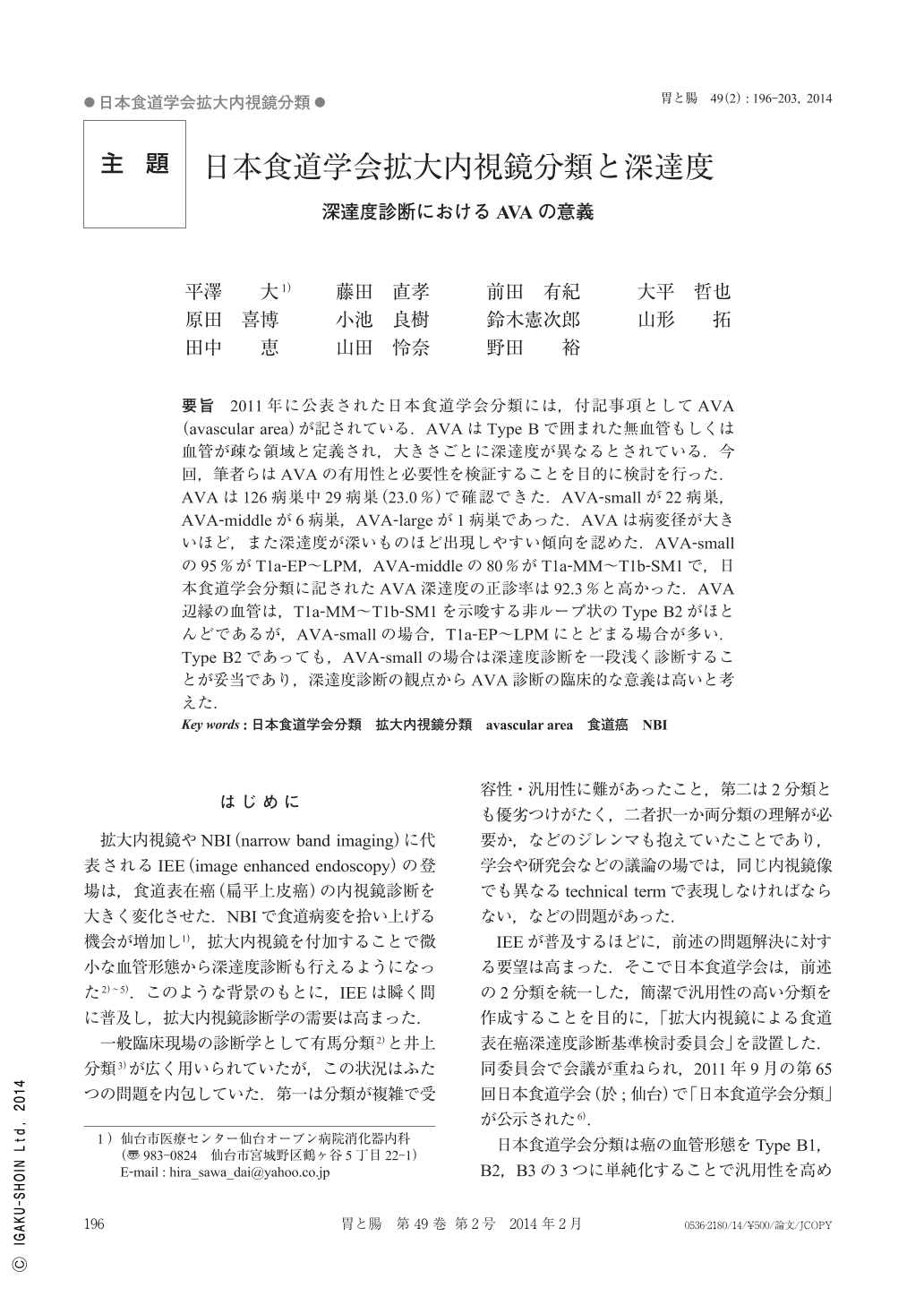
要旨 2011年に公表された日本食道学会分類には,付記事項としてAVA(avascular area)が記されている.AVAはType Bで囲まれた無血管もしくは血管が疎な領域と定義され,大きさごとに深達度が異なるとされている.今回,筆者らはAVAの有用性と必要性を検証することを目的に検討を行った.AVAは126病巣中29病巣(23.0%)で確認できた.AVA-smallが22病巣,AVA-middleが6病巣,AVA-largeが1病巣であった.AVAは病変径が大きいほど,また深達度が深いものほど出現しやすい傾向を認めた.AVA-smallの95%がT1a-EP~LPM,AVA-middleの80%がT1a-MM~T1b-SM1で,日本食道学会分類に記されたAVA深達度の正診率は92.3%と高かった.AVA辺縁の血管は,T1a-MM~T1b-SM1を示唆する非ループ状のType B2がほとんどであるが,AVA-smallの場合,T1a-EP~LPMにとどまる場合が多い.Type B2であっても,AVA-smallの場合は深達度診断を一段浅く診断することが妥当であり,深達度診断の観点からAVA診断の臨床的な意義は高いと考えた.
The AVA(avascular area)is described in the JES(Japan Esophageal Society)classification published in 2011 as a supplementary item. AVA is defined as an enclosed type B vessel and an area with sparse vessels with the invasion depth differing according to the size. We conducted a study to verify the usefulness and necessity of the AVA. The AVA was confirmed in 97 of 126 foci(23.0%). The size of the AVA was small in 20 foci, middle in 5 foci, and large in 1 focus. The AVA tended to be more easily recognized when tumor size was larger and invasion depth was deeper. The tumor reached the epithelium or lamina propria mucosa(T1a-EP-LPM)in 95% of small AVAs and the muscularis mucosa and submucosa up to 0.2mm(T1a-MM-T1b-SM1)in 80% of the middle AVAs. The accuracy of the AVA invasion depth described in the JES classification is thus high at 92.3%. Most of the vessels at the AVA margin were type B2 and non-loop-shaped, suggesting T1a-MM-T1b-SM1, but small AVAs usually remained at T1a-EP-LPM. It is appropriate to diagnose the invasion depth as shallow if the AVA is small, even if it is type B2. Furthermore, AVA diagnosis may have great clinical significance from the perspective of diagnosing the invasion depth through the follow-up period.

Copyright © 2014, Igaku-Shoin Ltd. All rights reserved.


