Japanese
English
- 有料閲覧
PPV購入案内
1,320円 (1,200円+税10%) こちらは、1201ページから1209ページの文献です。購入には医書.jp本会員登録が必要です。
会員登録完了後に改めて上記「購入サイトへ」を選択してください。
または「購入サイトへ」選択後に「購入ログイン」、「新規会員登録」の順に進んでください。
会員登録について詳しくはをご確認ください。
- Abstract 文献概要
- 1ページ目 Look Inside
- 参考文献 Reference
- サイト内被引用 Cited by
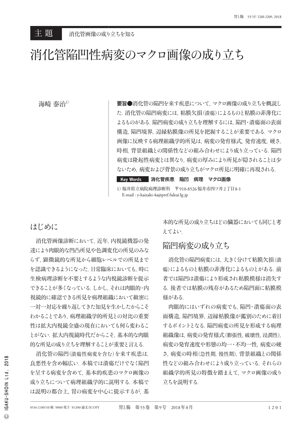
要旨●消化管の陥凹を来す疾患について,マクロ画像の成り立ちを概説した.消化管の陥凹病変には,粘膜欠損(潰瘍)によるものと粘膜の菲薄化によるものがある.陥凹病変の成り立ちを理解するには,陥凹・潰瘍面の表面構造,陥凹境界,辺縁粘膜像の所見を把握することが重要である.マクロ画像に反映する病理組織学的所見は,病変の発育様式,発育速度,硬さ,時相,背景組織との関係性などの組み合わせにより成り立っている.陥凹病変は隆起性病変とは異なり,病変の厚みにより所見が隠されることは少ないため,病変および背景の成り立ちがマクロ所見に明確に再現される.
In the present study, we outlined the principles of macroscopic imaging for depressed lesions of the gastrointestinal tract, which include those caused by mucosal defects(ulcers)and mucosal thinning. It is important to interpret the significance of the characteristics of the ulcerated surface, depressed boundary, and marginal mucosa to understand the formation of depressed lesions. Histological findings reflected in macroscopic images comprise a combination of the developmental pattern, developmental speed, rigidity, and time phase of the lesion and the relationship of the lesion with the background tissue. Unlike protruded lesions, depressed lesions are rarely hidden by the lesion thickness. Thus, the depressed lesions and the background tissue are clearly identifiable in macroscopic findings.
Copyright © 2018, Igaku-Shoin Ltd. All rights reserved.