Japanese
English
- 有料閲覧
PPV購入案内
1,320円 (1,200円+税10%) こちらは、440ページから449ページの文献です。購入には医書.jp本会員登録が必要です。
会員登録完了後に改めて上記「購入サイトへ」を選択してください。
または「購入サイトへ」選択後に「購入ログイン」、「新規会員登録」の順に進んでください。
会員登録について詳しくはをご確認ください。
- Abstract 文献概要
- 1ページ目 Look Inside
- 参考文献 Reference
- サイト内被引用 Cited by
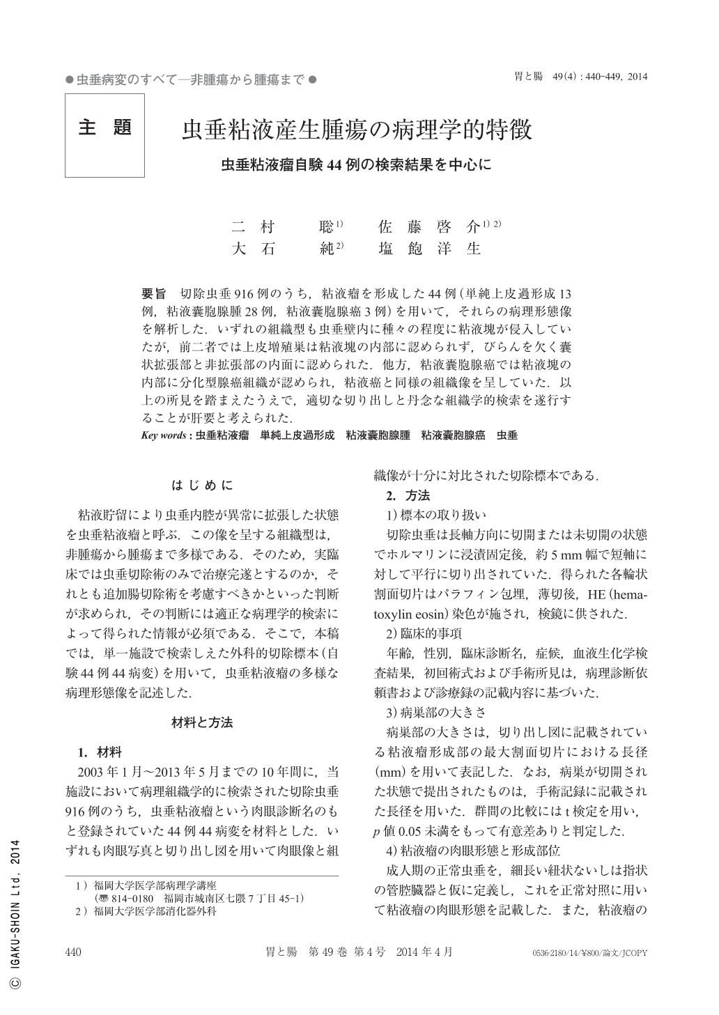
要旨 切除虫垂916例のうち,粘液瘤を形成した44例(単純上皮過形成13例,粘液囊胞腺腫28例,粘液囊胞腺癌3例)を用いて,それらの病理形態像を解析した.いずれの組織型も虫垂壁内に種々の程度に粘液塊が侵入していたが,前二者では上皮増殖巣は粘液塊の内部に認められず,びらんを欠く囊状拡張部と非拡張部の内面に認められた.他方,粘液囊胞腺癌では粘液塊の内部に分化型腺癌組織が認められ,粘液癌と同様の組織像を呈していた.以上の所見を踏まえたうえで,適切な切り出しと丹念な組織学的検索を遂行することが肝要と考えられた.
The aim of this study was to clarify the pathological features of appendiceal mucinous neoplasms. We examined 44 lesions from 44 cases with appendiceal mucocele(simple epithelial hyperplasia, mucinous cystadenoma, and mucinous cystadenocarcinoma : 13, 28, and 3 lesions, respectively)between January 2003 and May 2013 at our laboratory.
The following results were obtained. In all histologic types, penetration by mucus was also observed. Mucus devoid of epithelial cells was seen in the appendiceal wall, especially in histologic type of simple epithelial hyperplasia and mucinous cystadenoma. In sharp contrast, mucinous cystadenocarcinoma showed invasion of the appendiceal wall by irregular-shaped muconodules containing clearly identifiable differentiated adenocarcinoma components. This feature resembled that of colonic mucinous adenocarcinoma.
We conclude that appropriate cutting the resected specimens and carefully examination are essential to histological diagnosis of the appendiceal mucocele.
Copyright © 2014, Igaku-Shoin Ltd. All rights reserved.