Japanese
English
- 有料閲覧
PPV購入案内
1,320円 (1,200円+税10%) こちらは、499ページから510ページの文献です。購入には医書.jp本会員登録が必要です。
会員登録完了後に改めて上記「購入サイトへ」を選択してください。
または「購入サイトへ」選択後に「購入ログイン」、「新規会員登録」の順に進んでください。
会員登録について詳しくはをご確認ください。
- Abstract 文献概要
- 1ページ目 Look Inside
- 参考文献 Reference
- サイト内被引用 Cited by
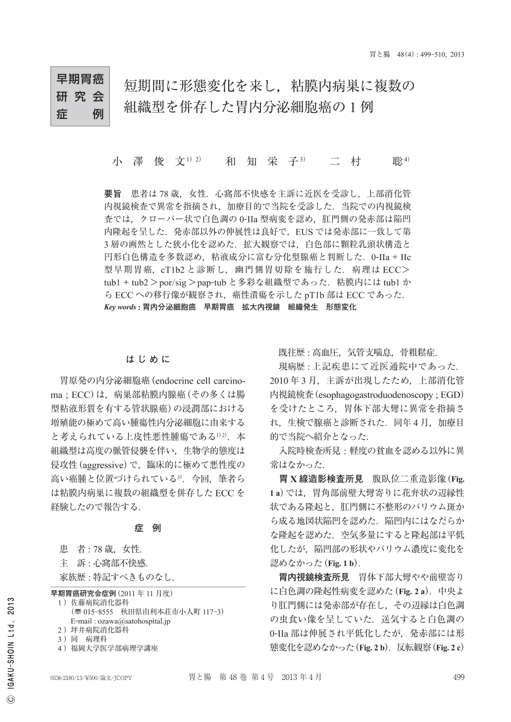
要旨 患者は78歳,女性.心窩部不快感を主訴に近医を受診し,上部消化管内視鏡検査で異常を指摘され,加療目的で当院を受診した.当院での内視鏡検査では,クローバー状で白色調の0-IIa型病変を認め,肛門側の発赤部は陥凹内隆起を呈した.発赤部以外の伸展性は良好で,EUSでは発赤部に一致して第3層の画然とした狭小化を認めた.拡大観察では,白色部に顆粒乳頭状構造と円形白色構造を多数認め,粘液成分に富む分化型腺癌と判断した.0-IIa+IIc型早期胃癌,cT1b2と診断し,幽門側胃切除を施行した.病理はECC>tub1+tub2>por/sig>pap-tubと多彩な組織型であった.粘膜内にはtub1からECCへの移行像が観察され,癌性潰瘍を示したpT1b部はECCであった.
A 78-year-old female visited our hospital because of an abnormality found during the ESD(esophago-gastro-duodenoscopy)at another clinic for epigastric discomfort. X-ray images showed a type 0-IIa+IIc lesion at the greater curvature of the lower gastric body. EGD showed a type 0-IIa whitish lesion in the form of clover-type, and a reddish depression with irregular margin in the distal portion. at the edge of this depression, we recognized a protrusion. Extensibility of this lesion was good except for the depressive area, and EUS(endoscopic ultrasonography)showed distinct narrowing of the third layer to match the depression. Magnifing endoscopy showed numerous white circular structures in the granular and papillary structure of the whitish part, so well-differentiated adenocarcinoma involving a mucinous component was presumed. Clinical diagnosis was early gastric cancer(type 0-IIa+IIc, with depth of invasion estimated as cT1b2), so the patient underwent distal gastrectomy. Pathological diagnosis was ECC(endocrine cell carcinoma)>tub1+tub2>por/sig>pap-tub. A variety of histological types of cancer were observed. In the mucosal layer, the transitional images of ECC from tub1 was observed, and showed the cancerous ulcer and part of the submucosal invasion was constituted by ECC.
Copyright © 2013, Igaku-Shoin Ltd. All rights reserved.