Japanese
English
- 有料閲覧
- Abstract 文献概要
- 1ページ目 Look Inside
- 参考文献 Reference
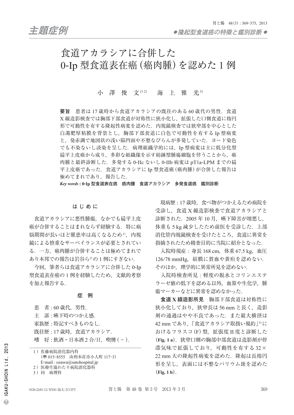
要旨 患者は17歳時から食道アカラシアの既往のある60歳代の男性.食道X線造影検査では胸部下部食道が対称性に狭小化し,拡張した口側食道に楕円形で可動性を有する隆起性病変を認めた.内視鏡検査では狭窄部を中心とした白濁肥厚粘膜を背景とし,胸部下部食道に白色で可動性を有するIp型病変と,発赤調で地図状の浅い陥凹面や不整なびらんが多発していた.ヨード染色でも不染ないし淡染を呈した.病理組織学的には,Ip型病変は主に低分化型扁平上皮癌から成り,多彩な組織像を示す紡錘型腫瘍細胞を伴うことから,癌肉腫と最終診断した.多発する0-IIcないし0-IIb病変はpT1a-LPMまでの扁平上皮癌であった.食道アカラシアにIp型食道癌(癌肉腫)が合併した報告は極めてまれであり,報告した.
A 61-year-old man with dysphagia was hospitalized. On esophagography, a symmetrical stenosis about 70mm in length was found in the lower esophagus. On the proximal side of the stenosis the protrusion was movabile. On esophagoscopy, a type 0-Ip protrusion with white coating and stalk located in the lower portion of the esophagus was noted. Furtheremore, multiple depressed lesions(0-IIc and 0-IIb), which had irregular shapes, as well as erosions were seen throughout the esophagus. On chromoendoscopy using Iodine staining, multiple lugol-voiding lesions were observed. An esophagectomy with lymphnode dissection was carried out. On histology, the 0-Ip lesion was composed of poorly differentiated squamous carcinoma cells and various spindle cell carcinoma cells, so a diagnosis of carcinosarcoma was made. Typical histological findings of achalasia were observed at the stenosis. An association of carcinosarcoma as a type 0-Ip lesion with esophageal achalasia is very rare.

Copyright © 2013, Igaku-Shoin Ltd. All rights reserved.


