Japanese
English
- 有料閲覧
PPV購入案内
1,320円 (1,200円+税10%) こちらは、571ページから578ページの文献です。購入には医書.jp本会員登録が必要です。
会員登録完了後に改めて上記「購入サイトへ」を選択してください。
または「購入サイトへ」選択後に「購入ログイン」、「新規会員登録」の順に進んでください。
会員登録について詳しくはをご確認ください。
- Abstract 文献概要
- 1ページ目 Look Inside
- 参考文献 Reference
- サイト内被引用 Cited by
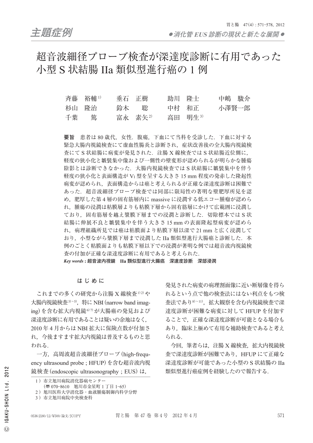
要旨 患者は80歳代,女性.腹痛,下血にて当科を受診した.下血に対する緊急大腸内視鏡検査にて虚血性腸炎と診断され,症状改善後の全大腸内視鏡検査にてS状結腸に病変が発見された.注腸X線検査ではS状結腸近位側に,軽度の狭小化と皺襞集中像および一側性の壁変形が認められるが明らかな腫瘍陰影とは診断できなかった.大腸内視鏡検査ではS状結腸に皺襞集中を伴う軽度の狭小化と表面構造がVI型を呈する大きさ15mm程度の発赤した隆起性病変が認められ,表面構造からは癌と考えられるが正確な深達度診断は困難であった.超音波細径プローブ検査では同部に限局性の著明な壁肥厚所見を認め,肥厚した第4層の固有筋層内にmassiveに浸潤する低エコー腫瘤が認められ,腫瘍の浸潤は粘膜層よりも粘膜下層から固有筋層にかけて広範囲に浸潤しており,固有筋層を越え漿膜下層までの浸潤と診断した.切除標本ではS状結腸に伸展不良と皺襞集中を伴う大きさ15mmの表面隆起型病変が認められ,病理組織所見では癌は粘膜面より粘膜下層以深で21mmと広く浸潤しており,小型ながら漿膜下層まで浸潤したIIa類似型進行大腸癌と診断した.本例のごとく粘膜面よりも粘膜下層以下での浸潤が著明な例では超音波内視鏡検査の付加が正確な深達度診断に有用であると考えられた.
An 80-year-old female was admitted with hematochezia and abdominal clamp. She was colonoscopically diagnosed as ischemic colitis. After recovery from her symptoms, total colonoscopy was carried out and a lesion was found. Though some stenosis with converging folds and unilateral deformity were delineated by barium enema study in the proximal sigmoid colon, definite diagnosis as a carcinoma was difficult. Colonoscopy revealed a 15mm sized of reddish flat elevated lesion and some stenosis with converging folds in the sigmoid colon. The lesion was diagnosed as a tubular adenocarcinoma by Vi pit surface pattern using magnifying colonoscopy, but invasion depth diagnosis could not be clearly made. HFUP revealed localized eminent wall thickness and a low echoic mass had extensively invaded massively beyond the SM layer into the proper muscle layer, and had partially invaded into the subserosal layer. The lesion was diagnosed as a small IIa-like advanced carcinoma. Histopathological diagnosis was a IIa-like advanced carcinoma(15mm in size in the mucosa and 21mm in size in submucosal layer, a moderately differentiated adenocarcinoma with invasion depth to the subserosa, ly1, v0, n1)as the preoperative HFUP diagnosis. We think that additional HFUP procedure is useful for the precise diagnosis of the invasion depth of a carcinoma with extensive invasion of the submucosal layer or deeper than the mucosal layer, as was the case in this patient.
Copyright © 2012, Igaku-Shoin Ltd. All rights reserved.