Japanese
English
- 有料閲覧
PPV購入案内
1,320円 (1,200円+税10%) こちらは、843ページから846ページの文献です。購入には医書.jp本会員登録が必要です。
会員登録完了後に改めて上記「購入サイトへ」を選択してください。
または「購入サイトへ」選択後に「購入ログイン」、「新規会員登録」の順に進んでください。
会員登録について詳しくはをご確認ください。
- Abstract 文献概要
- 1ページ目 Look Inside
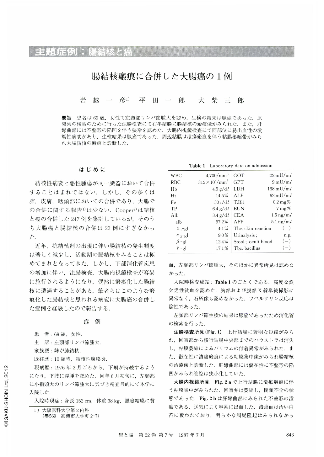
要旨 患者は69歳,女性で左頸部リンパ節腫大を認め,生検の結果は腺癌であった.原発巣の検索のために行った注腸検査にて右半結腸に腸結核の瘢痕像がみられた.また,肝彎曲部には不整形の陥凹を伴う狭窄を認めた.大腸内視鏡検査にて同部位に易出血性の潰瘍性病変があり,生検結果は腺癌であった.周辺粘膜は潰瘍瘢痕を伴う粘膜萎縮帯がみられ大腸結核の瘢痕と診断した.
The patient was a 69 year-old female who was admitted to our hospital complaining of swelling of a lymph node in the left neck. Resected specimen of the lymph node revealed adenocarcinoma. Therefore, examination of gastrointestinal tract was performed in order to detect the primary lesion. Upper gastrointestinal series showed no abnormalities. In the barium enema examination, marked shortening of the ascending colon was noted. Haustra was absent in the ascending colon and the proximal transverse colon. Convergence of the mucosal folds owing to the ulcer scar was scattered. In addition, stenosis of the lumen and irregular form of excavation were found in the hepatic flexure. In the colonofiberscopic examination, convergence of the mucosal folds in the ascending colon and incompetent ileocecal valve were observed. Irregular form of ulceration with dirty whitish coating was found in the hepatic flexure. Biopsy specimen taken from the ulcerated area revealed adenocarcinoma. Macroscopic finding of the resected specimen was type 2 of colonic carcinoma. Ascending colon was shortened and convergence of the mucosal folds was scattered. Histologically, well differentiated adenocarcinoma was found in the hepatic flexure. In the ascending colon, mild atrophy of the mucosa, interruption of the muscularis mucosae, and fibrosis in the submucosa were present. From these findings, diagnosis of colonic carcinoma complicated with tuberculous scar was made, though typical tuberculous tubercle was not found.
Copyright © 1987, Igaku-Shoin Ltd. All rights reserved.