Japanese
English
- 有料閲覧
- Abstract 文献概要
- 1ページ目 Look Inside
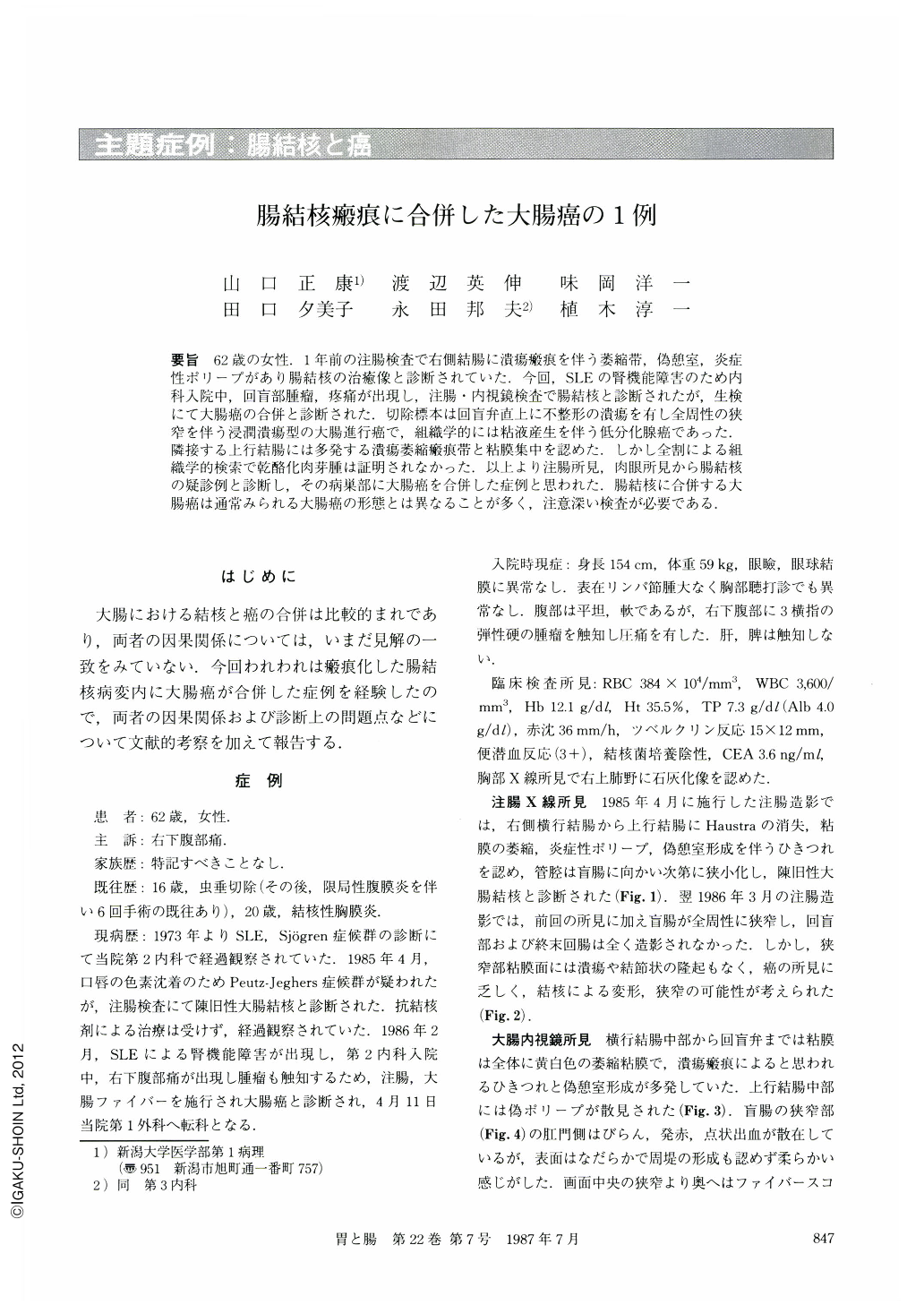
要旨 62歳の女性.1年前の注腸検査で右側結腸に潰瘍瘢痕を伴う萎縮帯,偽憩室,炎症性ポリープがあり腸結核の治癒像と診断されていた.今回,SLEの腎機能障害のため内科入院中,回盲部腫瘤,疼痛が出現し,注腸・内視鏡検査で腸結核と診断されたが,生検にて大腸癌の合併と診断された.切除標本は回盲弁直上に不整形の潰瘍を有し全周性の狭窄を伴う浸潤潰瘍型の大腸進行癌で,組織学的には粘液産生を伴う低分化腺癌であった.隣接する上行結腸には多発する潰瘍萎縮瘢痕帯と粘膜集中を認めた.しかし全割による組織学的検索で乾酪化肉芽腫は証明されなかった.以上より注腸所見,肉眼所見から腸結核の疑診例と診断し,その病巣部に大腸癌を合併した症例と思われた.腸結核に合併する大腸癌は通常みられる大腸癌の形態とは異なることが多く,注意深い検査が必要である.
A 62 year-old woman with right lower abdominal pain as her chief complaint was admitted to our hospital for detailed examination of the colon. One year before she had been diagnosed (by barium enema study) as having healed colon tuberculosis.
On the barium enema examination at the time of admission to our hospital, haustra was absent in the ascending colon. Convergence of the mucosal folds brought about by the ulcer scar was noticed here and there. In addition, stenosis of the lumen in the cecum was found.
In the colonofiberscopic examination, pseudodiverticula, multiple scars, and inflammatory polyps in the ascending colon were observed. A marked narrowing region with discolored, friable mucosa in the cecum was also observed. Biopsy taken from this narrowing region revealed adenocarcinoma. Thus, radiological and endoscopic examinations led to a diagnosis of colonic cancer accompanied with healed tuberculosis.
Macroscopic findings of the resected specimen revealed infiltrative ulcerating type of colonic carcinoma measuring 6×7cm in the ileo-cecal region. Convergence of the mucosal folds, inflammatory polyps and pseudodiverticula were scattered in the atrophic mucosa of the ascending colon.
Histologically, poorly to moderately differentiated adenocarcinoma with mucus products was found from the cecum to the terminal ileum. Cancer cells infiltrated scirrhously through the whole layers of the colon, invading the surrounding adipose tissues. Atrophic mucosa in the colon and ileum showed multiple shallow scars with fibromusculosis.

Copyright © 1987, Igaku-Shoin Ltd. All rights reserved.


