Japanese
English
- 有料閲覧
- Abstract 文献概要
- 1ページ目 Look Inside
- サイト内被引用 Cited by
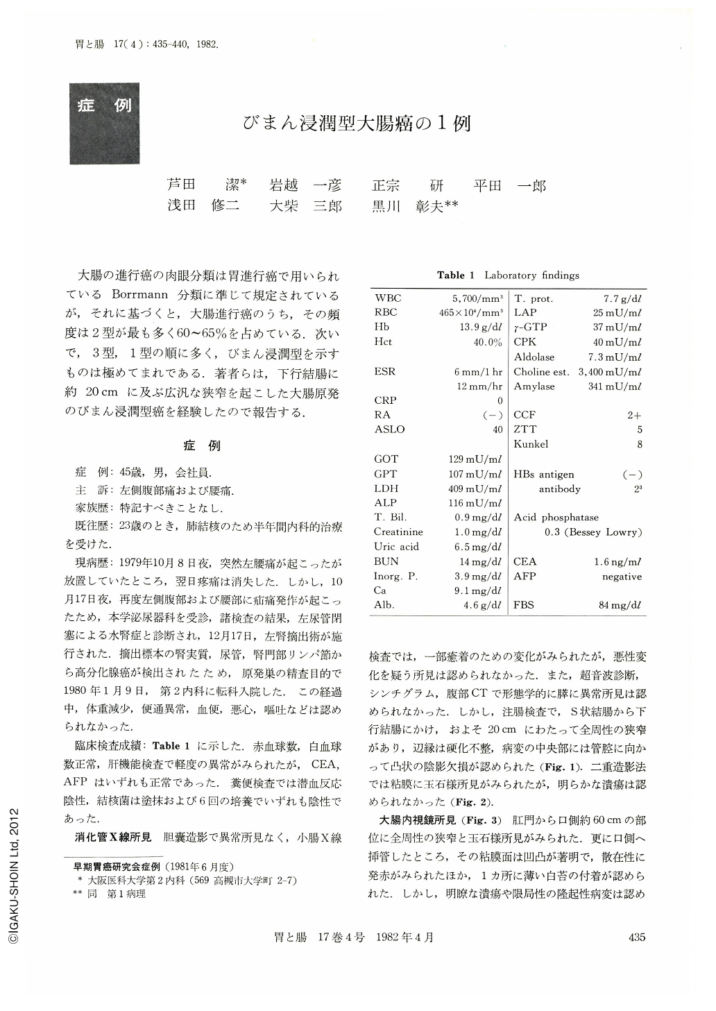
大腸の進行癌の肉眼分類は胃進行癌で用いられているBorrmann分類に準じて規定されているが,それに基づくと,大腸進行癌のうち,その頻度は2型が最も多く60~65%を占めている.次いで,3型,1型の順に多く,びまん浸潤型を示すものは極めてまれである.著者らは,下行結腸に約20cmに及ぶ広汎な狭窄を起こした大腸原発のびまん浸潤型癌を経験したので報告する.
This report presents a case of diffusely infiltrating type of colonic carcinoma. The patient was a 45-year-old man who was admitted to the Department of Urology complaining of left-side flank pain and lumbago. As well-differentiated adenocarcinoma was found from the specimens of the left kidney, ureter and lymph nodes taken when left nephrectomy was performed, he was referred to our Department to detect primary lesion. Upper G-I series and cholecystogram showed no abnormalities. Barium enema examination demonstrated circumferential narrowing in the distal descending colon and a part of the sigmoid colon. No clear-cut demarcation was observed between the lesion and intact colon. The colonic wall was rigid and irregular. In the middle of the lesion, filling defect was demonstrated. Cobblestone appearance in the lesion was shown in the double contrast method. In the endoscopic examination, narrowing of the lumen and cobblestone appearance were demonstrated 60 cm proximal from the anus. Irregular mucosa accompanied by redness was observed by deeper insertion of the scope. Biopsy specimen taken from the lesion showed well-differentiated adenocarcinoma. A part of the sigmoid colon and the distal descending colon were resected.
Macroscopic findings of the resected specimen were as follows: In the middle of the resected specimen, the lumen showed belt-shaped narrowing. The wall of this area was somewhat thickened. A part of the mucosa of this area was protuberant. The mucosa apart from this area was apparently intact. Histologically, only in the small part of the central lesion, adenocarcinoma with well-differentiated type was exposed over the mucosal surface. In the remaining part, carcinoma cells existed only in the submucosa and muscular layer and surface was covered by normal colonic mucosa. In the proximal and distal area apart from the lesion, infiltration into the lymph vessels of carcinoma cells in the submucosa, muscular layer and serosa associated with fibrosis was predominant. Mucus was stained by PAS or Alcian blue in the cytoplasma of the carcinoma cells. From these findings, diagnosis of diffusely infiltrating type of the primary colonic carcinoma was confirmed.
Copyright © 1982, Igaku-Shoin Ltd. All rights reserved.