Japanese
English
- 有料閲覧
- Abstract 文献概要
- 1ページ目 Look Inside
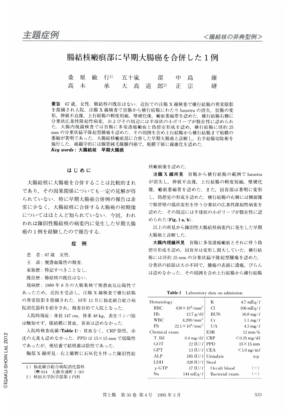
要旨 67歳,女性.肺結核の既往はない.近医での注腸X線検査で横行結腸の異常陰影を指摘され入院.注腸X線検査で盲腸から横行結腸にわたりhaustraの消失,盲腸の変形,伸展不良像,上行結腸の軽度短縮,壁硬化像,瘢痕萎縮帯を認めた.横行結腸右側に分葉状広基性隆起性病変,およびその周辺には半球状の小ポリープが散在性に認められた.大腸内視鏡検査では盲腸に多発潰瘍瘢痕と偽憩室形成を認め,横行結腸に径約25mmの分葉状扁平隆起型腫瘍を認めた.その周囲を含め上行結腸から横行結腸まで粘膜の萎縮が著明であった.大腸結核瘢痕部に合併した早期大腸癌と診断し,右半結腸切除術を施行した.組織学的には腺管絨毛腺腫内癌で,粘膜下層に線維化を認めた.
A 65-year-old woman was admitted to our hospital for further evaluation of positive indication of occult blood in her stool. Barium enema examination showed ahaustral appearance and marked shortening from the ascending colon to the proximal transverse colon. Nodule-aggregating lesion (2 cm in diamiter) was also observed in the proximal transverse colon. Colonoscopic findings showed multiple ulcer scars, pseudodiverticula and atrophic mucosa caused by tuberculous colitis. A superficial-elevated type early colonic cancer was demonstrated in the proximal trasverse colon. These findings led us to a diagnosis of early colonic cancer occurring at the site of healed tuberculous colitis. Microscopic findings of the resected specimen revealed mucosal carcinoma with tubulovillous adenoma in the area of healed tuberculous colitis.

Copyright © 1995, Igaku-Shoin Ltd. All rights reserved.


