Japanese
English
- 有料閲覧
PPV購入案内
1,320円 (1,200円+税10%) こちらは、1057ページから1061ページの文献です。購入には医書.jp本会員登録が必要です。
会員登録完了後に改めて上記「購入サイトへ」を選択してください。
または「購入サイトへ」選択後に「購入ログイン」、「新規会員登録」の順に進んでください。
会員登録について詳しくはをご確認ください。
- Abstract 文献概要
- 1ページ目 Look Inside
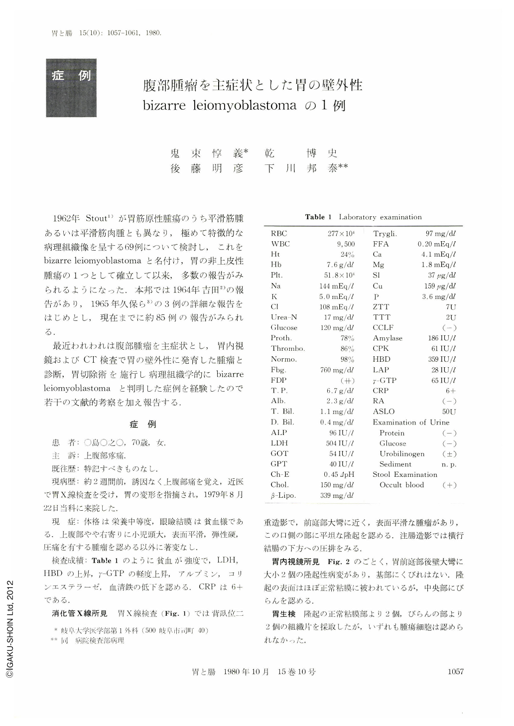
1962年Stout1)が胃筋原性腫瘍のうち平滑筋腫あるいは平滑筋肉腫とも異なり,極めて特徴的な病理組織像を呈する69例について検討し,これをbizarre leiomyoblastomaと名付け,胃の非上皮性腫瘍の1つとして確立して以来,多数の報告がみられるようになった.本邦では1964年吉田2)の報告があり,1965年久保ら3)の3例の詳細な報告をはじめとし,現在までに約85例の報告がみられる.
最近われわれは腹部腫瘤を主症状とし,胃内視鏡およびCT検査で胃の壁外性に発育した腫瘤と診断,胃切除術を施行し病理組織学的にbizarre leiomyoblastomaと判明した症例を経験したので若干の文献的考察を加え報告する.
A 70 year-old woman was admitted to the hospital on August 22, 1979. with a complaint of upper abdominal pain. On physical examination the conjunctiva was anemic. A large, elastic hard mass was palpable, filling the right upper abdomen. The mass was tender and fixed.
An upper gastrointestinal roentgenologic examination demonstrated two protruded lesions of the antrum. With gastrofiberscopy, the lesions were found to be covered by the smooth mucosa with erosion on the top of the tumor. Biopsy specimens obtained from the surface of the tumor and the margin of the erosion showed no neoplastic cells. Selective celiac angiogram showed slight pooling of the dye on the greater curvature of the antrum in capillary phase. CT showed a large low density mass having high density margin and septums in the anterior right abdomen. The mass had a gas shadow near the anterior abdominal wall, which was suspected of the stomach gas. The margin and the septums of the tumor were enhanced by infusion of angiographin.
Subtotal gastrectomy with transverse colectomy was performed. Gross findings of the resected stomach showed an extraluminally growing, 15×12×8 cm tumor on the greater curvature of the antrum. Cut sections through the tumor showed marked lobulation with many friable cystic areas containing a hamorrhagic purulent material. Microscopically, round or polygonal cells with clear zones around the nucleus were seen. Based on these microscopic findings, a diagnosis of bizarre leiomyoblastoma was established.
Copyright © 1980, Igaku-Shoin Ltd. All rights reserved.