Japanese
English
- 有料閲覧
PPV購入案内
1,320円 (1,200円+税10%) こちらは、1075ページから1079ページの文献です。購入には医書.jp本会員登録が必要です。
会員登録完了後に改めて上記「購入サイトへ」を選択してください。
または「購入サイトへ」選択後に「購入ログイン」、「新規会員登録」の順に進んでください。
会員登録について詳しくはをご確認ください。
- Abstract 文献概要
- 1ページ目 Look Inside
主として壁外性に増殖した空腸の平滑筋肉腫の症例を報告する.
A 46year-old man complained slight pain on the left lower abdomen in the night of August 16, 1980. On August 17, he had hematochezia three times. On August 18, he had a large amount of melena, palpitation, vertigo and cold sweating. Laboratory findings showed RBC 219×104/ml, Hb 7.1g/dl, Occult Blood Uuaiac (+++), WBC 9,500, Total-Protein 5.0g/dl and others were within normal limits.
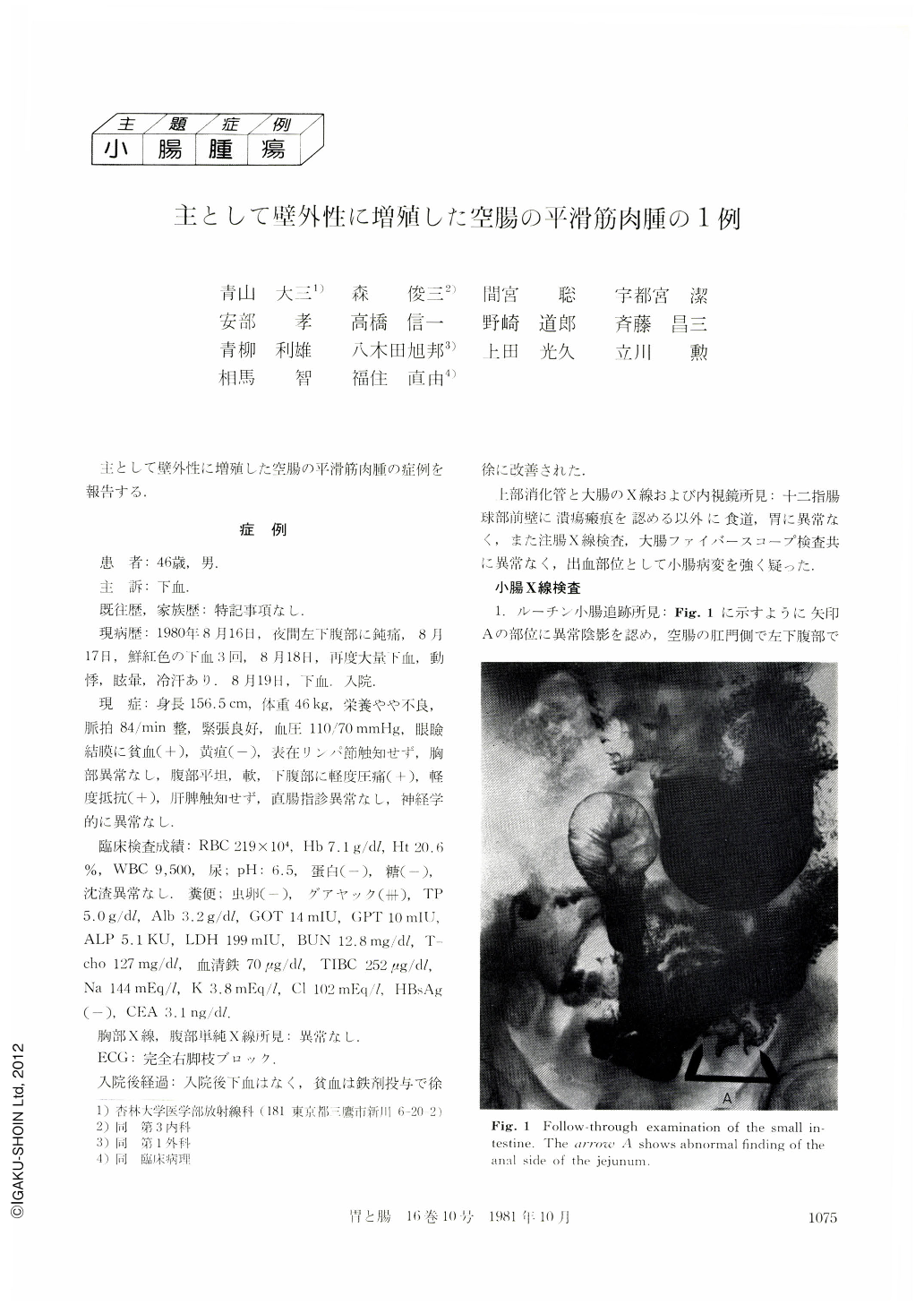
X-ray examinations of chest, abdomen, upper G-I and lower G-I showed no particular abnormal findings. Fig. 1 shows the follow-through radiogram of the small intestine and arrow"A"shows the abnormal findings of the distal jejunum. Fig. 2 demonstrates the compressions-method radiogram, and Fig 4 shows the double contrast radiogram. Fig. 6 and 7 show the angiographic findings. At a glance, Fig. 2 and Fig. 4 seem to indicate"leiomyomatous tumor", however, the finer findings are not quite understandable.
The operation disclosed the extraluminal tumor of 5×6×3cm at the distal site of the jejunum, 150cm from the Treitz's band, and no metastasis was found anywhere. Histological finding was leiomyosarcoma.
From the retrospecti veviews of the resected specimen, the radiological findings were very confusing. The irregularities of the jejunum wall showing in “C” of Fig. 3 and 5 are due to the fat lesion surrounding the tumor. The contour of the tumor is shown in “E” of the Fig. 3 and 5. The intraluminal small (17×12mm) prominence with the central small (5×2mm) depression are well demonstrated in “D” of the Fig. 3 and 5.
The radiological findings of the extraluminal proliferating tumor of the small intestine are complicated and sometimes it could be misdiagnosed. Therefore, the combined methods of compression, double-contrast and angiographic method should be employed, however instead of those, the diagnosis would be still very difficult.
Copyright © 1981, Igaku-Shoin Ltd. All rights reserved.