Japanese
English
- 有料閲覧
PPV購入案内
1,320円 (1,200円+税10%) こちらは、1209ページから1212ページの文献です。購入には医書.jp本会員登録が必要です。
会員登録完了後に改めて上記「購入サイトへ」を選択してください。
または「購入サイトへ」選択後に「購入ログイン」、「新規会員登録」の順に進んでください。
会員登録について詳しくはをご確認ください。
- Abstract 文献概要
- 1ページ目 Look Inside
症 例
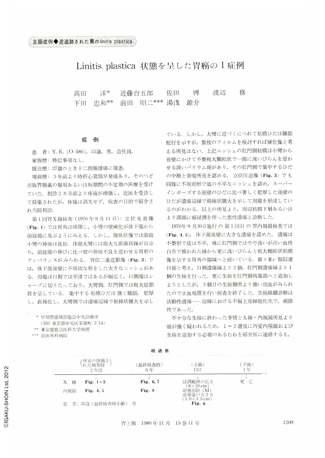
患 者:Y. K.(O-586),33歳,男,会社員.
家族歴:特記事項なし.
既往歴:27歳のとき十二指腸潰瘍に罹患.
現病歴:3年前より時折心窩部早発痛あり,そのつど市販胃腸薬の服用あるいは短期間の不定期の医療を受けていた.初診2カ月前より疼痛が増強し,近医を受診して投薬されたが,疼痛は消失せず,検査の目的で紹介され当院初診.
A 33 year-old man complanied of epigastric pain. The initial radiological and gastroscopic examinations (Sept. 1976) revealed abnormality resembling early gastric cancer type Ⅲ+Ⅱc on the posterior wall of lower body (presumptive width of original lesion: 30×35 mm), that is, irregularly-shaped giant ulcer niche with uneven bottom surface and surrounding mucosal pattern with shallow depression at the anal side of the ulcer margin and thick converging folds toward oral and greater curvature of the ulcer.
At this time it was diagnosed as advanced cancer of the stomach having cancerous infiltration to the deeper layer suspiciously. Then biopsy was performed at the oral and anal margin of the ulcer (three specimens), but the following biopsy had to stop because of bleeding. Unfortunately, the histological diagnosis of pioptimized specimen was no malignancy. And so re-biopsy was recommended, but the patient refused it, and he never came to our clinic. When he returned after two years, his complaints were worse than his initial visit.
The final radiograph and gastroscopy (Sept. 1978) revealed extreme decrease in distensibility of the stomach wall as well as coarse, irregularly-winded and stiff mucosal rugae at the region from subcardiac to angular portion and easily diagnosed as gastric carcinoma (linitis plastica type).
Surgical operation was carried out on Sept. 29, 1978. The pathological diagnosis of resected specimen was adenocarcinoma scirrhosum, poorly differenciated with serosal cancerous infiltration and positive lymph nodes metastasis. The width of the cancerous lesion was 8×10 cm. There are still many unsolved problems to be faced in making early diagnosis of gastric carcinoma scirrhous type, but the most important clue to the detection of these scirrhous carcinoma of the stomach in its early stage is to check for cancerous erosion accompanied with ulcerous lesion.
Copyright © 1980, Igaku-Shoin Ltd. All rights reserved.