Japanese
English
- 有料閲覧
- Abstract 文献概要
- 1ページ目 Look Inside
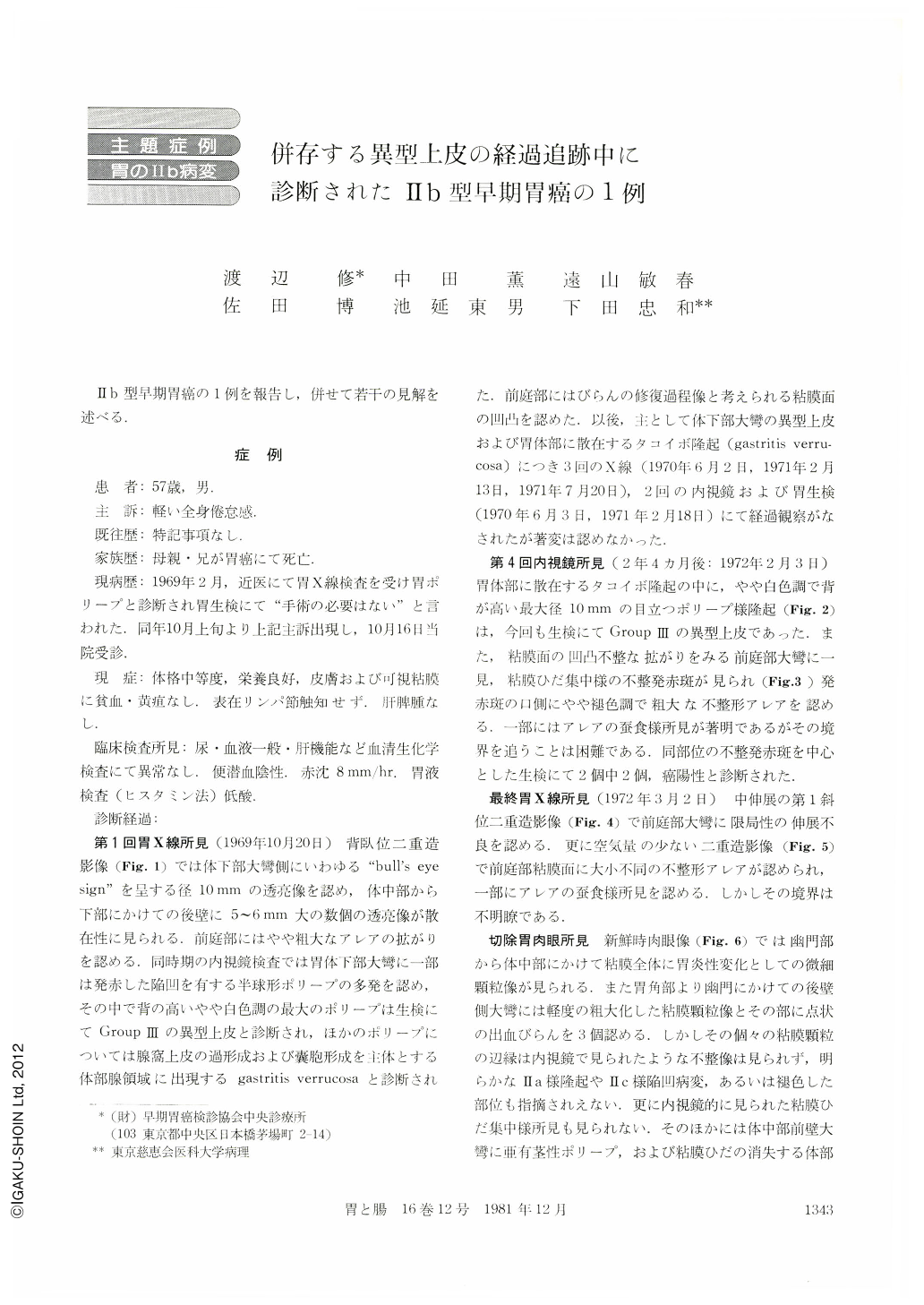
Ⅱb型早期胃癌の1例を報告し,併せて若干の見解を述べる.
A 57-year-old man visited our clinic complaining of slight general fatigue in Feb. 1969. By radiological and gastroendoscopic examinations, scattered hemispherical polypoid lesions (gastritis verrucosa) and a tall, whitish polypoid lesion, diagnosed as atypical epithelium (group Ⅲ) by biopsy specimens, were found on the corpus of his stomach. After that, the follow-up studies of the atypical epithelium and gastritis verrucosa were made by radiological, gastroendoscopic and bioptic examinations, but no significant changes were observed. At the fourth gastroendoscopic examination (28 months after the first exam.), irregularly-shaped and reddish depression was found in the rough and irregular mucosal surface of the antrum. It seemed like some converging folds. And slightly-whitish, coarse and irregular“area gastricae”were seen in the oral side of the depression. Some parts of these“area gastricae”showed distinct wormeaten appearances in outline. Cancer cells were found in the part of depression by biopsy.
On the double contrast films of the last radiological examination, we were able to indicate limited, descreased distension on the greater curvature of antrum. Besides, we were able to detect some worm-eaten appearance, at a few parts of “area gastricae”on the antrum, but the border of abnormal area was not lined out clearly.
In macroscopic picture of the resected stomach granular mucosal surface patterns were seen on both antrum and corpus. Slightly coarse, granular mucosa with three hemorrhagic erosions were also shown in the posterior wall of antrum.
However, neither Ⅱa-like elevation non Ⅱc-like depression was found in this area. In microscopic findings, the cancer cell's invasion was limited mainly within the mucosal propria at the posterior wall of antrum.
The size of cancer region was 40×35mm, and histological diagnosis was poorly differentiated tubular adenocarcinoma. Its cross sections also showed almost flat surface between the region invaded by cancer and the surrounding normal mucosa.
Therefore, this case was considered as a typical Ⅱb-type early gastric cancer.

Copyright © 1981, Igaku-Shoin Ltd. All rights reserved.


