Japanese
English
- 有料閲覧
PPV購入案内
1,320円 (1,200円+税10%) こちらは、21ページから26ページの文献です。購入には医書.jp本会員登録が必要です。
会員登録完了後に改めて上記「購入サイトへ」を選択してください。
または「購入サイトへ」選択後に「購入ログイン」、「新規会員登録」の順に進んでください。
会員登録について詳しくはをご確認ください。
- Abstract 文献概要
- 1ページ目 Look Inside
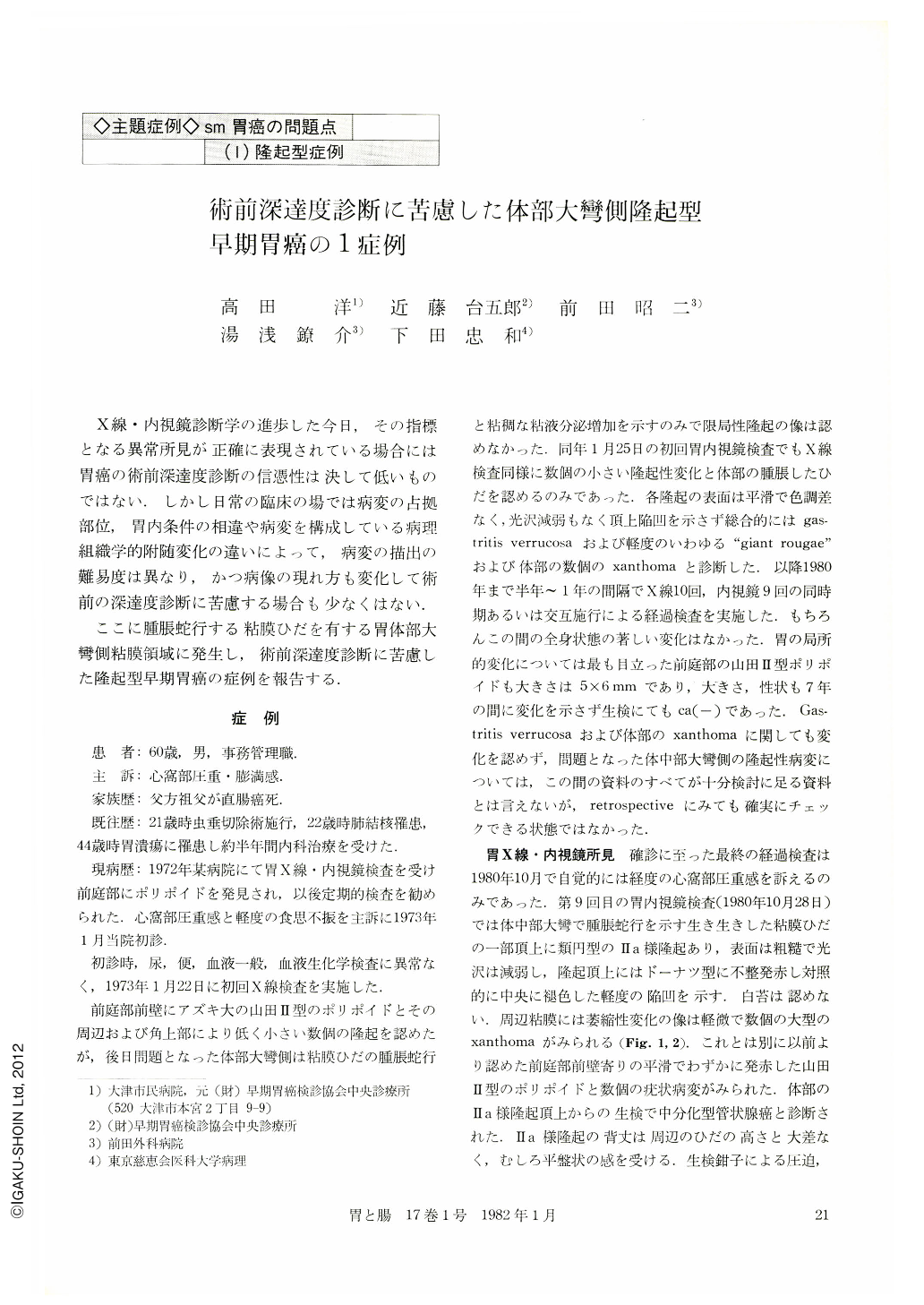
X線・内視鏡診断学の進歩した今日,その指標となる異常所見が正確に表現されている場合には胃癌の術前深達度診断の信憑性は決して低いものではない.しかし日常の臨床の場では病変の占拠部位,胃内条件の相違や病変を構成している病理組織学的附随変化の違いによって,病変の描出の難易度は異なり,かつ病像の現れ方も変化して術前の深達度診断に苦慮する場合も少なくはない.
ここに腫脹蛇行する粘膜ひだを有する胃体部大彎側粘膜領域に発生し,術前深達度診断に苦慮した隆起型早期胃癌の症例を報告する.
An early gastric cancer lesion was found on the greater curvature of the gastric body in a 60-year-old man, who had been followed-up for seven years because of a polypoid lesion (eosinophilic granuloma) of the antrum.
On the x-ray and endoscopic examinations, the lesion was detected on the greater curvature of the middle of the body, about 12×10 mm in diameter, and was observed on the weaving swollen mucosal folds of the greater curvature.
The surface of the lesion was rough and lusterless, and showed doughnut-shaped redness with a central discoloration on the top of the lesion endoscopically. The lesion was most likely Ⅱa type early gastric cancer in which cancerous infiltration was suspected to be mucosal layer alone because of a lack of irregularity, rigidity and an apparent central ulceration on the lesion and its surrounding mucosal folds. Although one spot-film with a compression study on the x-ray examination demonstrated an apparent central depression on the top of the lesion, which might indicate a further invasion of cancer tissues to the submucosal layer, this radiological finding easily disappeared by intensifying the compression on the lesion.
The subsequent histology of the resected specimen demonstrated that cancerous infiltration reached partially to the submucosal layer, and the lesion was therefore classified into sm type early gastric cancer. The histological type of the lesion was a moderately differentiated adenocarcinoma, and a moderate edema and mild fibrosis were seen in submucosal layer, which could have formed an elevated lesion in this case.
The lesion on the greater curvature of the body might be misinterpreted in determination of depth of cancerous invasion on both x-ray and endoscopic examination because of weaving swollen folds. But if we had checked the central depression of this lesion more carefully on the x-ray examination we might have been able to get the correct diagnosis of the depth of cancerous invasion.
Copyright © 1982, Igaku-Shoin Ltd. All rights reserved.