Japanese
English
- 有料閲覧
PPV購入案内
1,320円 (1,200円+税10%) こちらは、957ページから961ページの文献です。購入には医書.jp本会員登録が必要です。
会員登録完了後に改めて上記「購入サイトへ」を選択してください。
または「購入サイトへ」選択後に「購入ログイン」、「新規会員登録」の順に進んでください。
会員登録について詳しくはをご確認ください。
- Abstract 文献概要
- 1ページ目 Look Inside
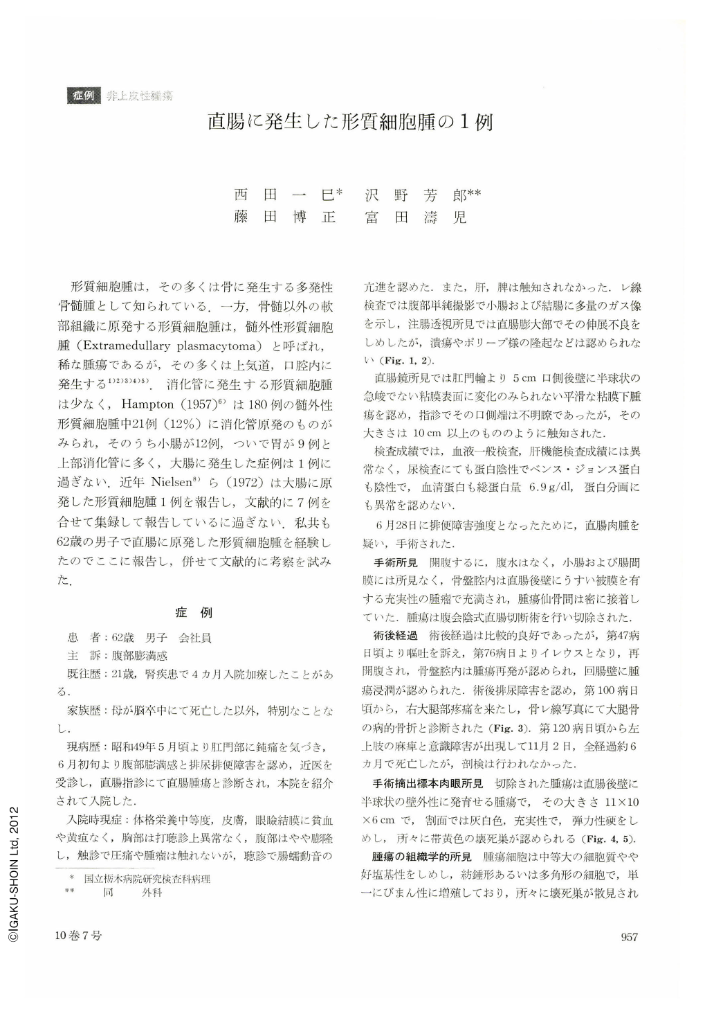
形質細胞腫は,その多くは骨に発生する多発性骨髄腫として知られている.一方,骨髄以外の軟部組織に原発する形質細胞腫は,髄外性形質細胞腫(Extramedullary plasmacytoma)と呼ばれ,稀な腫瘍であるが,その多くは上気道,口腔内に発生する1)2)3)4)5).消化管に発生する形質細胞腫は少なく,Hampton(1957)6)は180例の髄外性形質細胞腫中21例(12%)に消化管原発のものがみられ,そのうち小腸が12例,ついで胃が9例と上部消化管に多く,大腸に発生した症例は1例に過ぎない.近年Nielsen8)ら(1972)は大腸に原発した形質細胞腫1例を報告し,文献的に7例を合せて集録して報告しているに過ぎない.私共も62歳の男子で直腸に原発した形質細胞腫を経験したのでここに報告し,併せて文献的に考察を試みた.
The case: a white collar worker, male, 62 years of age.
Since about two months before he had a feeling of abdominal distention and defecation trouble. A doctor in his neighborhood made a diagnosis of rectal tumor. He was then referred to the surgical department of our hospital. Barium enema examination at admission revealed a marked stricture in the ampulla recti. Digital examination of the rectum together with rectoscopy confirmed a hemispheric submucosal tumor on the posterior wall 5 cm oral from the anal ring. Neither examination of the blood nor liver function tests revealed nothing of abnormal value. B.-J's protein was negative in the urine. Electrophoresis of the serum protein was also normal.
As the above findings led us to suspect rectal sarcoma, surgical operation was performed. The tumor was large enough, filling the pelvic cavity reaching from the posterior wall of the rectum to the sacrum. The tumor was resected including the rectum. Relaparotomy was later done because ileus developed about 76 days after the operation. Recurrence of the tumor was then detected in the ileum wall. The patient began to complain of pain in the right thigh, confirmed by X-ray as due to pathologic fracture of the right femur. He became worse day by day, dying about six months after the initial examination.
The resected tumor was a hemispheric tumor on the posterior wall of the rectum, showing extramural development. It measured 11×10×6 cm. Its cut surface was solid with grayish white color, at places showing yellow necrosis.
Histologically, the tumor was plasmacytoma with pleomorphic cells including giant cells.
Copyright © 1975, Igaku-Shoin Ltd. All rights reserved.