Japanese
English
- 有料閲覧
PPV購入案内
1,320円 (1,200円+税10%) こちらは、1185ページから1188ページの文献です。購入には医書.jp本会員登録が必要です。
会員登録完了後に改めて上記「購入サイトへ」を選択してください。
または「購入サイトへ」選択後に「購入ログイン」、「新規会員登録」の順に進んでください。
会員登録について詳しくはをご確認ください。
- Abstract 文献概要
- 1ページ目 Look Inside
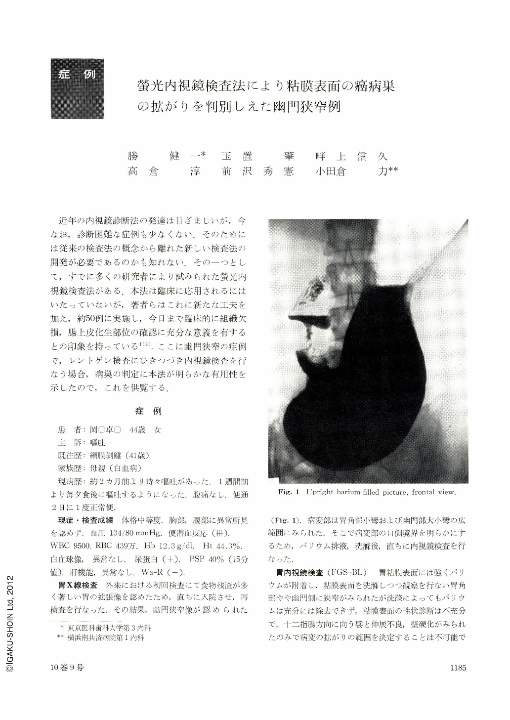
近年の内視鏡診断法の発達は目ざましいが,今なお,診断困難な症例も少なくない.そのためには従来の検査法の概念から離れた新しい検査法の開発が必要であるのかも知れない.その一つとして,すでに多くの研究者により試みられた螢光内視鏡検査法がある.本法は臨床に応用されるにはいたっていないが,著者らはこれに新たな工夫を加え,約50例に実施し,今日まで臨床的に組織欠損,腸上皮化生部位の確認に充分な意義を有するとの印象を持っている1)2).ここに幽門狭窄の症例で,レントゲン検査にひきつづき内視鏡検査を行なう場合,病巣の判定に本法が明らかな有用性を示したので,これを供覧する.
The patient, a 44-year-old female, was admitted because of severe vomiting at each meals for the previous one week. The upper G-I series examination revealed pyloric stenosis. Subsequent endoscopical examination revealed pyloric stenotic findings with constriction. Its mucosal surface was coated with barium meals. The margin of malignant lesion could not be found due to constriction of gastric wall and barium meals. Then, we performed fluorescein endoscopic examination, (Gastroenterological Endoscopy, 17(3): 36~40, 1975). The malignant lesion of the gastric mucosa was revealed to have red color region after 15 seconds of fluorescein injection from antecubital vein. The fluorescien endoscopic picture of this paitient revealed yellow green color on the normal mucosal surface, but the stenotic region was surrounded by red-colored mucosa. The patient underwent surgical treatment for the gastrectomy. On the resected stomach, 6.5×5.5 cm large round protruded tumor with crater was shown on the antrum. Histologically, the main lesion was adenocarcinoma mucocellulare combined with adenocarcinoma simplex, infiltrating into the serosa. This case presented a clinical features suggestive of the malignant tumor of the stomach but its exact extent of the malignant lesion was not determined by the conventional techniques for G-Ⅰ examinations. New method of fluorescein endoscopic examination proved to be useful for evaluation of malignant lesion of gastric surface. We concluded that this method is very useful to diagnose malignant lesion of the stomach and to determine its extent.
Copyright © 1975, Igaku-Shoin Ltd. All rights reserved.