Japanese
English
- 有料閲覧
PPV購入案内
1,320円 (1,200円+税10%) こちらは、925ページから930ページの文献です。購入には医書.jp本会員登録が必要です。
会員登録完了後に改めて上記「購入サイトへ」を選択してください。
または「購入サイトへ」選択後に「購入ログイン」、「新規会員登録」の順に進んでください。
会員登録について詳しくはをご確認ください。
- Abstract 文献概要
- 1ページ目 Look Inside
従来より胃の悪性リンパ腫については,全身性系統的に発生した悪性リンパ腫の一部分症として,胃に発生したものと,胃粘膜のリンパ細網組織より発生したいわゆる胃原発性悪性リンパ腫とは一応区別して考えられてきた.
臨床診断面では最近の医療器械の改良と診断技術の飛躍的向上にもかかわらず,胃の悪性リンパ腫は胃癌および胃潰瘍との鑑別,病理組織学的には未分化癌およびreactive lymphoreticular hyperplasia(RLH)などとの鑑別が問題とされ,発生頻度が少ないことと相まって,術前診断が困難な病変とされている,最近筆者らは胃X線,胃内視鏡,胃生検,生検細胞診,リンパ管造影などにより,胃原発性リンパ肉腫と診断しえた症例について報告する.
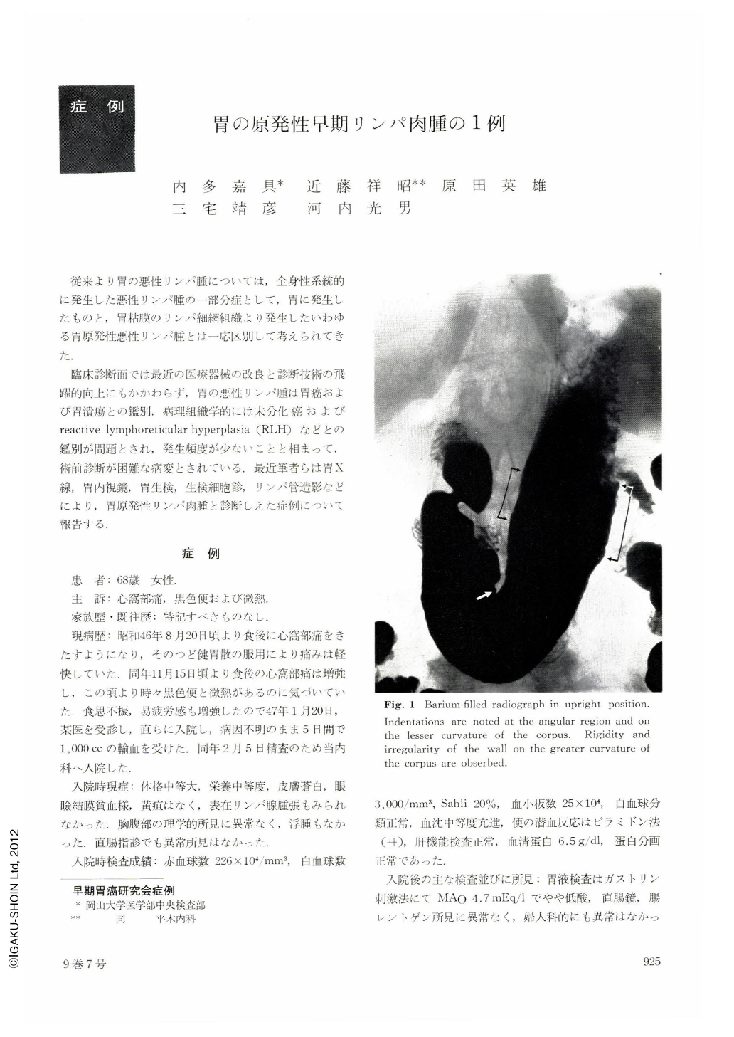
The patient, a 68-year-old woman, visited the department of internal medicine in the affiliated hospital in our medical school, complaining of epigastric pain, tarry stool and subfebrile temperature. Her chief complaints led us to suspect a disease in the upper GI tract and some hematological disorders, and examinations were accordingly performed. The peripheral blood picture was that of hypochromic anemia with no juvenile leucocytes. Examination of the bone marrow was normal. The initial roentgenography of the stomach revealed in an upright, barium-filled picture deformities of the lesser curvature in the antrum, of both curvatures in the body and the angle. In supine double contrast pictures were seen several protruding lesions of low stature on the posterior wall of the mid-body. Gastroscopy showed several low elevations in each of the lesions on the posterior wall of the body and on the lesser curvature of the antrum near the angle. Their surface was whitish, showing normal highlights. Neither reddening nor bleeding was observed. Both roentgenography and endoscopy of the stomach was hardly revealing in the distinction between carcinoma, RLH, or submucosal tumor. Cytology of the biopsied specimens with May-Giemsa staining showed a number of well-distributed cells, definitely different from those of the epithelium. They were judged lymphoblasts by their morphologic features. As an ancillary finding plasmocytes were also found, No phagocyte, polymorph or eosinophil were to be seen. Histologic examination of the biopsied specimens yielded much difficulty in the discrimination between cancer and malignant lymphoma even by the use of special staining method.
Finally we arrived at a diagnosis of lymphoblastic lymphoblarcoma after taking account of cytodiagnosis of biopsied materials. Histological study of the resected stomach was early multiple Lymphosarcoma of gastric origin.
Copyright © 1974, Igaku-Shoin Ltd. All rights reserved.