Japanese
English
- 有料閲覧
PPV購入案内
1,320円 (1,200円+税10%) こちらは、549ページから554ページの文献です。購入には医書.jp本会員登録が必要です。
会員登録完了後に改めて上記「購入サイトへ」を選択してください。
または「購入サイトへ」選択後に「購入ログイン」、「新規会員登録」の順に進んでください。
会員登録について詳しくはをご確認ください。
- Abstract 文献概要
- 1ページ目 Look Inside
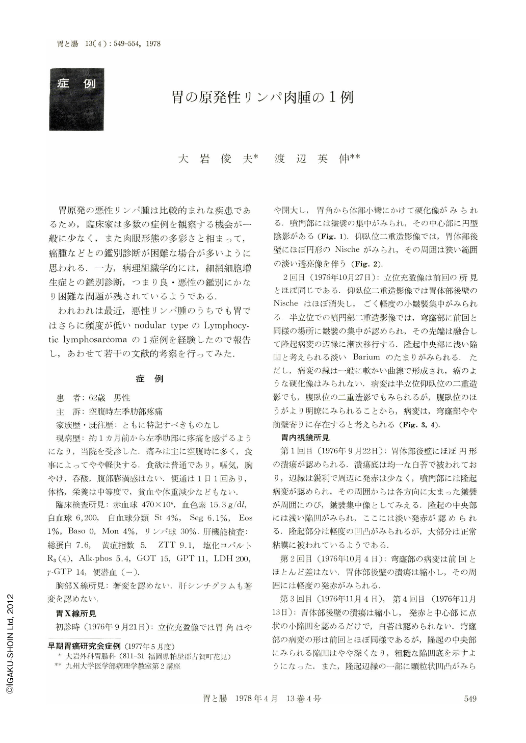
胃原発の悪性リンパ腫は比較的まれな疾患であるため,臨床家は多数の症例を観察する機会が一般に少なく,また肉眼形態の多彩さと相まって,癌腫などとの鑑別診断が困難な場合が多いように思われる.一方,病理組織学的には,細網細胞増生症との鑑別診断,つまり良・悪性の鑑別にかなり困難な問題が残されているようである.
われわれは最近,悪性リンパ腫のうちでも胃ではさらに頻度が低いnodular typeのLymphocytic lymphosarcomaの1症例を経験したので報告し,あわせて若干の文献的考察を行ってみた.
A sixty-two-year-old man visited Oiwa clinic with a complaint of hunger pain in the epigastric area. The pain had continued for a month. Upper GI tract series showed a well-defined elevated lesion in the fornix of the stomach. A shallow barium fleck was noticed in the middle of this elevated lesion and several large folds were also found extending to it. These folds looked like converging folds, but they flowed smoothly onto the elevated surface of the lesion. The entire lesion of tha area showed a smooth pattern without any evidence of the rigidity of cancer. A niche was also noticed on the posterior wall of the body.
Endoscopic examination revealed a nodular elevation with large folds in the fornix of the stomach. A shallow, red depression was observed in the middle of the lesion. The surface of the elevated lesion was irregular in shape and the entire lesion was covered with the same mucosa as the surrounding tissue. A small ulcer covered with white coating was also found in the posterior wall of the body.
With medical treatment for one month, the ulcer in the posterior wall of the body showed improvement. Biopsy specimens were taken three times from the elevated lesion in the fornix. Although the first two biopsy attempts proved unsuccessful, the third positively revealed it as lymphosarcoma.
Total gastrectomy was performed. The resected specimen showed a 2.5×2.5×1.5 cm sized tumor with a 1.8×1.7 cm sized erosion in the anterior wall of the fornix. located three cm from the esophagocardiac junction. A scar of the healed ulcer was also found in the posterior wall of the gastric body.
Histological study revealed an infiltration of matured lymphoma cells and was diagnosed as lymphocytic lymphosarcoma.
Copyright © 1978, Igaku-Shoin Ltd. All rights reserved.