Japanese
English
- 有料閲覧
PPV購入案内
1,320円 (1,200円+税10%) こちらは、1175ページから1181ページの文献です。購入には医書.jp本会員登録が必要です。
会員登録完了後に改めて上記「購入サイトへ」を選択してください。
または「購入サイトへ」選択後に「購入ログイン」、「新規会員登録」の順に進んでください。
会員登録について詳しくはをご確認ください。
- Abstract 文献概要
- 1ページ目 Look Inside
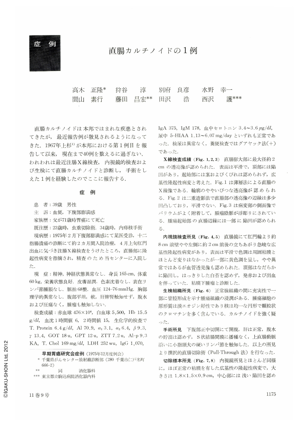
直腸カルチノイドは本邦ではまれな疾患とされてきたが,最近報告例が散見されるようになってきた.1967年上杉1)が本邦における第1例目を報告して以来,現在まで40例を数えるに過ぎない.われわれは最近注腸X線検査,内視鏡的検査および生検にて直腸カルチノイドと診断し,手術をしえた1例を経験したのでここに報告する.
The patient was 39-year-old male complaining of bloody stool and a sense of fullness in the lower abdomen.
Physical examination revealed no pathological findings. Clinical laboratory data were almost normal except for positive occult blood. The amount of serotonin in the blood and the excretion of 5-HIAA into urine were within normal limits.
A sessile elevated lesion with smooth surface measuring 2×2 cm was revealed roentgenographically on the anterior wall of the rectal ampulla. Endoscopically, a well-demarcated tumor was situated laterally and anteriorly about 8 cm from the anus. The tumor was mostly of normal color the same as the surrounding mucosa. Partially it was yellowish. Mucosal surface of the tumor was even, and a shallow depression with bleeding was noted on its tip. Biopsy specimen was diagnosed as carcinoid histologically.
Operation revealed the tumor measuring 1.8×1.5 cm in width and 0.9 cm in height with an erosion on its tip. The invasion of carcinoid was limited within the submucous layer. There was no metastatic liver tumor, although metastasis to regional lymph node was proved histologically. Silver staing was positive.
Rectal carcinoid is a rare disease and 40 cases including this one have been reported in Japan. Recent diagnostic methods such as double contrast study and endoscopic examination with biopsy will surely increase positive diagnostic rate of such a rare disorder of the gastrointestinal tract.
Copyright © 1976, Igaku-Shoin Ltd. All rights reserved.