Japanese
English
- 有料閲覧
PPV購入案内
1,320円 (1,200円+税10%) こちらは、1309ページから1313ページの文献です。購入には医書.jp本会員登録が必要です。
会員登録完了後に改めて上記「購入サイトへ」を選択してください。
または「購入サイトへ」選択後に「購入ログイン」、「新規会員登録」の順に進んでください。
会員登録について詳しくはをご確認ください。
- Abstract 文献概要
- 1ページ目 Look Inside
はじめに
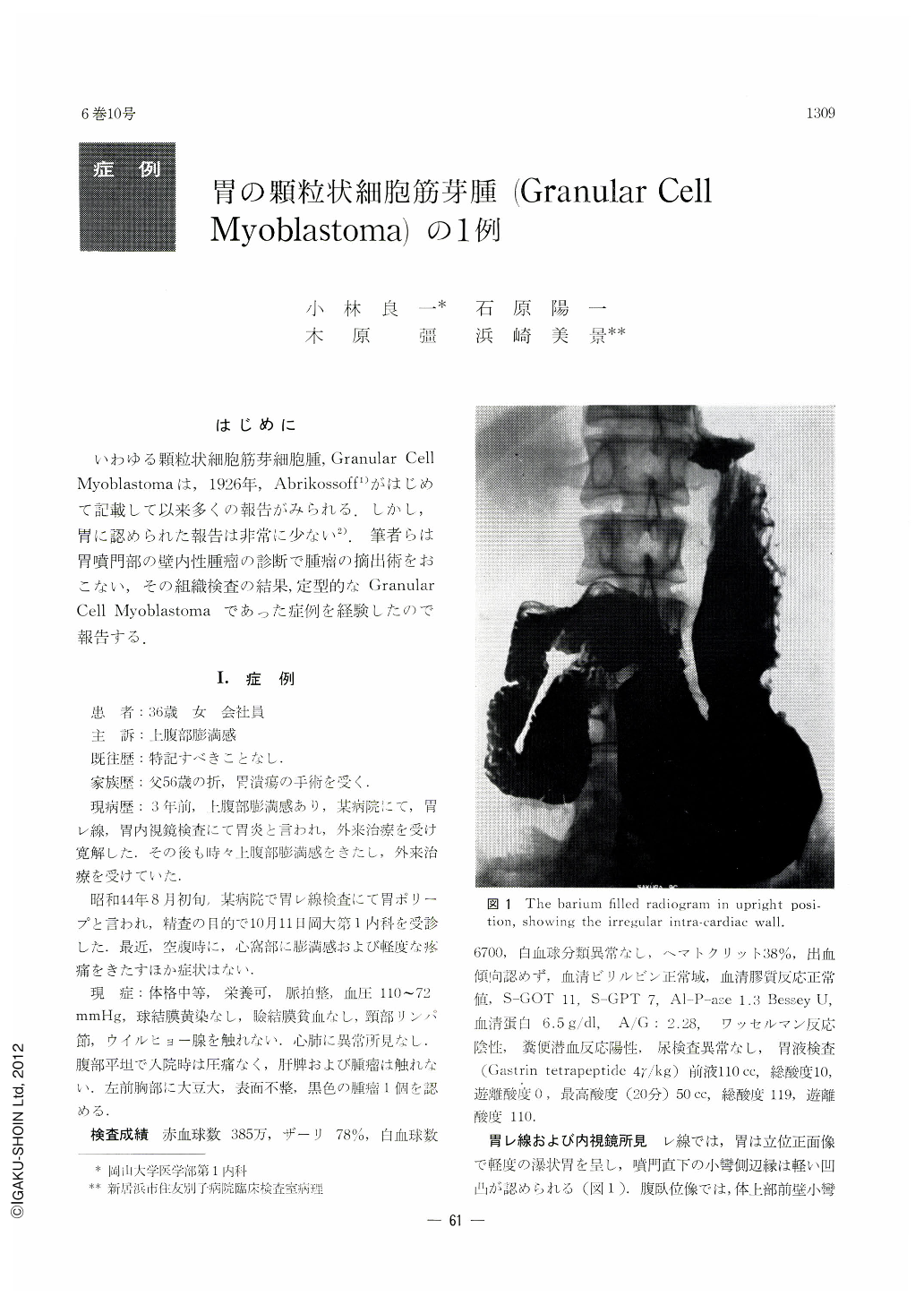
いわゆる顆粒状細胞筋芽細胞腫,Granular Cell Myoblastomaは,1926年,Abrikossoff1)がはじめて記載して以来多くの報告がみられる.しかし,胃に認められた報告は非常に少ない2).筆者らは胃噴門部の壁内性腫瘤の診断で腫瘤の摘出術をおこない,その組織検査の結果,定型的なGranular Cell Myoblastomaであった症例を経験したので報告する.
A 36-year-old woman was admitted to the Department of Internal Medicine, Okayama University Hospital with a suspicion of polyp of the stomach on November 4, 1969. She had been complaining of mid-epigastric fullness during the previous three years.
Physical and laboratory examinations proved normal except for slight anemia. X-ray study of the stomach revealed a small, well-defined filling defect on the anterior wall of the cardia in addition to ulcer on the angulus. Gastroscopic examination disclosed the former to be a small, hemispeheric tumor of smooth surface accompanied with a bridging fold over it. These findings were very suggestive of leiomyoma especially as its incidence is most high among intramural neoplasms of the stomach. Concomitant benign ulcer at the angulus, when treated for one month, disappeared, so that only the tumor was removed. It was a parenchymatous and yellow nodule of elastic and firm consistency, measuring 1.0×1.0×0.7 cm. Histologically it was confirmed as a granular cell myoblastoma.
It is a very rare neoplasm of the stomach and so far only several cases have been reported in the world literature. In Japan it is believed to be the second. The first (1965) was associated with neurilemmoma of the stomach.
Copyright © 1971, Igaku-Shoin Ltd. All rights reserved.