Japanese
English
- 有料閲覧
- Abstract 文献概要
- 1ページ目 Look Inside
はじめに
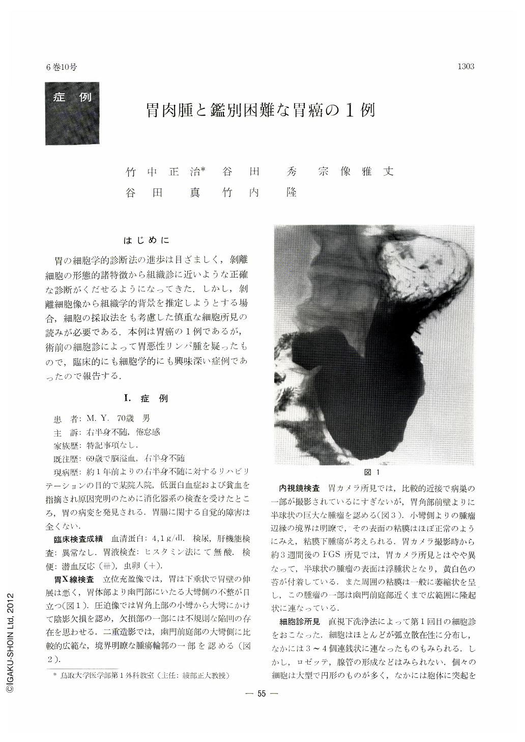
胃の細胞学的診断法の進歩は目ざましく,剝離細胞の形態的諸特徴から組織診に近いような正確な診断がくだせるようになってきた.しかし,剝離細胞像から組織学的背景を推定しようとする場合,細胞の採取法をも考慮した慎重な細胞所見の読みが必要である.本例は胃癌の1例であるが,術前の細胞診によって胃悪性リンパ腫を疑ったもので,臨床的にも細胞学的にも興味深い症例であったので報告する.
This paper deals with a case of gastric cancer that was very diificult to diagnose before the surgical operation even through various diagnostic procedures. Cytological examinations were done twice by lavage method. The exfoliated cells were larger in size, mostly solitary and dispersed. The cells were either round in shape or irregular with protruding cytoplasma, which showed deep basophilic staining. A clear halo was recognized around the nuclei and their chromatin was less fine and more granular. Mytosis was also seen in many of the cells. These cytological findings led the authors to suspect malignant lymphoma. Direct smear cytological examination after the operation showed distinct epithelial connection and finally this tumor was diagnosed as cancer. Histopathologically, it was undifferentiated adenocarcinoma.
Findings of the tumor cells in this cases afforded a very suggestive information regarding the differentiation between cancer and reticulum cell sarcoma. Grave reflection was also called for.
As compared with cancer of the stomach, its malignant lymphoma clinically shows better prognosis with more promising postoperative chemotherapy, so that correct diagnosis before the operation is all the more important, and because it is very diflicult to discriminate between cancer and malignant lymphoma only by endoscopy and x-ray, gastric biopsy and cytological examination are of great significance in this respect.

Copyright © 1971, Igaku-Shoin Ltd. All rights reserved.


