Japanese
English
- 有料閲覧
PPV購入案内
1,320円 (1,200円+税10%) こちらは、685ページから691ページの文献です。購入には医書.jp本会員登録が必要です。
会員登録完了後に改めて上記「購入サイトへ」を選択してください。
または「購入サイトへ」選択後に「購入ログイン」、「新規会員登録」の順に進んでください。
会員登録について詳しくはをご確認ください。
- Abstract 文献概要
- 1ページ目 Look Inside
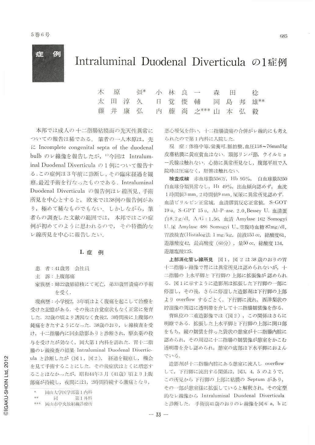
本邦では成人の十二指腸粘膜面の先天性異常についての報告は稀である.筆者の一人木原は,先にIncomplete congenital septa of the duodenal bulbのレ線像を報告したが,1)今回はIntraluminal Duodenal Diverticulaの1例について報告する.この症例は3年前に診断し,その臨床経過を観察,最近手術を行なったものである.Intraluminal Duodenal Diverticula報告例はレ線所見,手術所見を中心とすると,欧米では38例の報告例があり,極めて稀なものでもない.しかしながら,筆者らの調査した文献の範囲では,本邦ではこの症例が初めてのように思われるので,その特徴的なレ線所見を中心に報告したい.
Intraluminal duodenal diverticulum, formerly considered as of rare occurrence, has been reported with increasing frequency since communications of Schmidt (1941), Nelson (1947) and Kinzer (1949). To the best of our knowledge, 38 cases of this anomaly have been described in America and Europe, but never in Japan.
This is a report of a intraluminal duodenal diveticulum including its typical radiographic features and operative findings encountered in 38-yearoid man who had been complaining of epigastric pain for several years. An upper gastrointestinal x-ray series (Figs. 1 and 2) demonstrated a peculiar defect extending from the mid-descending duodenum into the pars horizontalis. As he had continued symptoms in spite of medical management, he underwent surgical exploration on May 22, 1969.
Pathognomonic roentgen features of intraluminal diverticulum are demonstrated in Figs. 1 and 2. X-ray picture shows a smooth, pouch or pear-like barium-filled structure within the lumen of the bowel (Figs. 3, 4 and 5) The wall or sac of the diverticulum is readily outlined as a thin translucent zone 1~2 mm thick surrounding the diverticulum except at the base of its attachment, which is found in the descending limb of the duodenum (Figs. 6 and 7).
The duodenum was mobilized by Kocher's maneuver and opened. The diverticulum was immediately apparent resembling a finger of a glove (Fig. 8). It was related cosely at its origin to the supra-ampullary region (Fig. 9). Simple excision of the diverticulum at its base was carried out and duodenal ulcer being associated, gastrectomy and gastroduodenostomy were performed as well.
The postoperative course was uneventful. He was discharged symptom-free on July 7, 1969.
The histologic appearance of the diverticulum is that of normal duodenal mucosa lining both sides of the sac. A small amount of incomplete muscularis mucosae and fibrous tissue is seen (Fig. 10 and 11).
Copyright © 1970, Igaku-Shoin Ltd. All rights reserved.