Japanese
English
- 有料閲覧
- Abstract 文献概要
- 1ページ目 Look Inside
はじめに
現在では,早期胃癌の診断は日常茶飯事のことになりっっあるが,そのなかでとくにⅡb型早期胃癌の診断はまだ難かしい点が多い.筆者らは,最近内視鏡検査で異常な発赤としてとらえられ,生検で癌と診断し得た4×4mmの微小Ⅱbを経験したので報告する.この症例は病理肉眼像でも病変を指摘することは困難であり,現在の胃癌診断の限界を示すと思われる.この症例が今後のⅡb型早期胃癌に対する診断の糸口ともなり,癌発生問題への一助となることを期待している.
A case of small Ⅱb type early gastric cancer diagnosed by aiming biopsy.
An early gastric cancer, measuring only 4 by 4 mm, was preoperatively diagnosed as such by biopsy. The lesion was first found in the course of FGS examination under direct view as an abnormal engorgement among manifestations of atrophic gastritis. This case was of interest because early gastric cancer of Ⅱb type as compared with its other varieties still harbors many difficulties in the diagnosis.
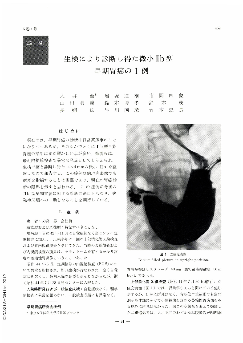
A 60-year-old man was found to have an abnormality in his stomach when in June 1969 he underwent a periodical gastric examination by endoscopy (FGS) followed by biopsy on the same day. On 28 of the following month he was admitted to the author's Center for further study. X-ray pictures of the upper gastrointestinal tract done after his admission showed only slight round protrusions of various sizes all over the pyloric antrum even after careful examination by changing not only the amount of air in his stomach but also that of the contrast medium. Roentgenologically it was a picture of atrophic gastritis. Besides extensive atrophic changes as seen in June, endoscopy with FGS disclosed among them a xanthoma on the greater curvature of the antrum, and on its aboral side was seen a protrusion slightly engorged. The lesion was inconspicuous among all the other changes, although it could be pointed out as an abnormality in the pictures of GTF-A. Evans' blue method showed clearly a picture of gastritis with specific intestinal metaplasia appearing as engroged protrusion on the distal side of the xanthoma slightly larger than other protrusions. Of 2 specimens taken by biopsy in June, cancer was diagnosed in the first one, and operation was performed in August. Gross finging of the resected stomach in fresh, half-fixed and fixed specimens hardly gave hint of cancer lesion though atrophic gastritis and xanthoma on the greater curvature of the antrum were evident. Histologically, the lesion was adenocarcinoma tubulare, mearuing only 4×4 mm, localized within the mucosal layer.

Copyright © 1970, Igaku-Shoin Ltd. All rights reserved.


