Japanese
English
- 有料閲覧
PPV購入案内
1,320円 (1,200円+税10%) こちらは、659ページから682ページの文献です。購入には医書.jp本会員登録が必要です。
会員登録完了後に改めて上記「購入サイトへ」を選択してください。
または「購入サイトへ」選択後に「購入ログイン」、「新規会員登録」の順に進んでください。
会員登録について詳しくはをご確認ください。
- Abstract 文献概要
- 1ページ目 Look Inside
- サイト内被引用 Cited by
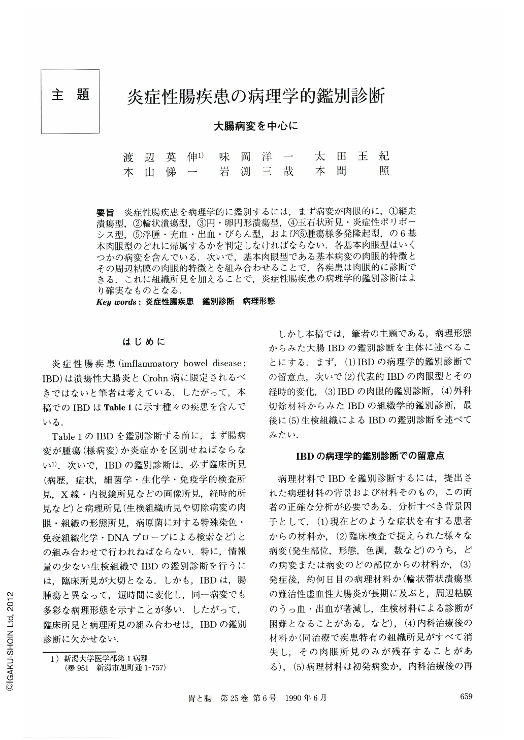
要旨 炎症性腸疾患を病理学的に鑑別するには,まず病変が肉眼的に,①縦走潰瘍型,②輪状潰瘍型,③円・卵円形潰瘍型,④玉石状所見・炎症性ポリポーシス型,⑤浮腫・充血・出血・びらん型,および⑥腫瘍様多発隆起型,の6基本肉眼型のどれに帰属するかを判定しなければならない.各基本肉眼型はいくつかの病変を含んでいる.次いで,基本肉眼型である基本病変の肉眼的特徴とその周辺粘膜の肉眼的特徴とを組み合わせることで,各疾患は肉眼的に診断できる.これに組織所見を加えることで,炎症性腸疾患の病理学的鑑別診断はより確実なものとなる.
In the diagnosis of inflammatory bowel diseases (IBD) which involve the small and/or large intestines, macroscopic findings are more useful than microscopic ones. Although microscopic changes sometimes completely disappear spontaneously or after medical treatment, macroscopic changes continue to exist.
The first key-step to the diagnosis is to classify main macroscopic findings into one of the six basic types (Table 3): 1) longitudinal ulcer, 2) circular ulcer, 3) round or oval ulcer, 4) cobblestone appearance and/or inflammatory polyposis, 5) edema, hyperemia, hemorrhage and erosions, and 6) tumor-like polypoid lesions.
Each basic macroscopic type can derive from a variety of diseases as shown in the right column of Table 3. These diseases are macroscopically differentiated each other according to the second key-step, which is to take into consideration the changes of the mucosa (Tables 6~11) surrounding the main macroscopic lesions.
It is possible to make a correct macroscopic diagnosis with an accuracy of about 90% according to these two key-steps and even with higher accuracy by combining histological findings.
Copyright © 1990, Igaku-Shoin Ltd. All rights reserved.