Japanese
English
- 有料閲覧
PPV購入案内
1,320円 (1,200円+税10%) こちらは、479ページから484ページの文献です。購入には医書.jp本会員登録が必要です。
会員登録完了後に改めて上記「購入サイトへ」を選択してください。
または「購入サイトへ」選択後に「購入ログイン」、「新規会員登録」の順に進んでください。
会員登録について詳しくはをご確認ください。
- Abstract 文献概要
- 1ページ目 Look Inside
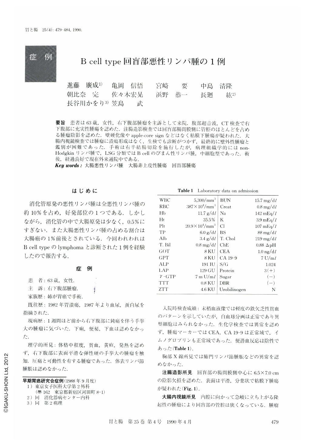
要旨 患者は63歳,女性,右下腹部腫瘤を主訴として来院.腹部超音波,CT検査で右下腹部に充実性腫瘍を認めた.注腸造影検査では回盲部腸間膜側に管腔のほとんどを占める腫瘤陰影を認めた.壁硬化像やapple-core signなどはなく粘膜下腫瘍が疑われた.大腸内視鏡検査では腫瘤に潰瘍形成はなく,生検でも診断がつかず,最終的に壁外性腫瘤と鑑別が困難であった.手術は右半結腸切除を施行したが,病理組織学的にはnon-Hodgkinリンパ腫で,LSG分類ではB cellのびまん性リンパ腫,中細胞型であった.術後,経過良好で現在外来通院中である.
A 63-year-old woman was admitted to our hospital because of abdominal mass. Ultrasonography was used as a primary screening procedure on this patient with a palpable mass in the right lower quadrant. It showed a solid mass in the RLQ. Barium enema showed a filling defect of the cecum. Colonoscopy when carefully observed revealed a submucosal tumor in the cecum. Examination of the biopsy specimen, however, did not lead to a specific diagnosis. Curative right hemicolectomy was performed on June 4, 1988. Histologically the tumor consisted of non-Hodgkin lymphoma, diffuse, medium-sized cell type.
Postoperative chemotherapy was done and the patient has been well as of the time of writing this manuscript.
Copyright © 1990, Igaku-Shoin Ltd. All rights reserved.