Japanese
English
- 有料閲覧
PPV購入案内
1,320円 (1,200円+税10%) こちらは、555ページから557ページの文献です。購入には医書.jp本会員登録が必要です。
会員登録完了後に改めて上記「購入サイトへ」を選択してください。
または「購入サイトへ」選択後に「購入ログイン」、「新規会員登録」の順に進んでください。
会員登録について詳しくはをご確認ください。
- Abstract 文献概要
- 1ページ目 Look Inside
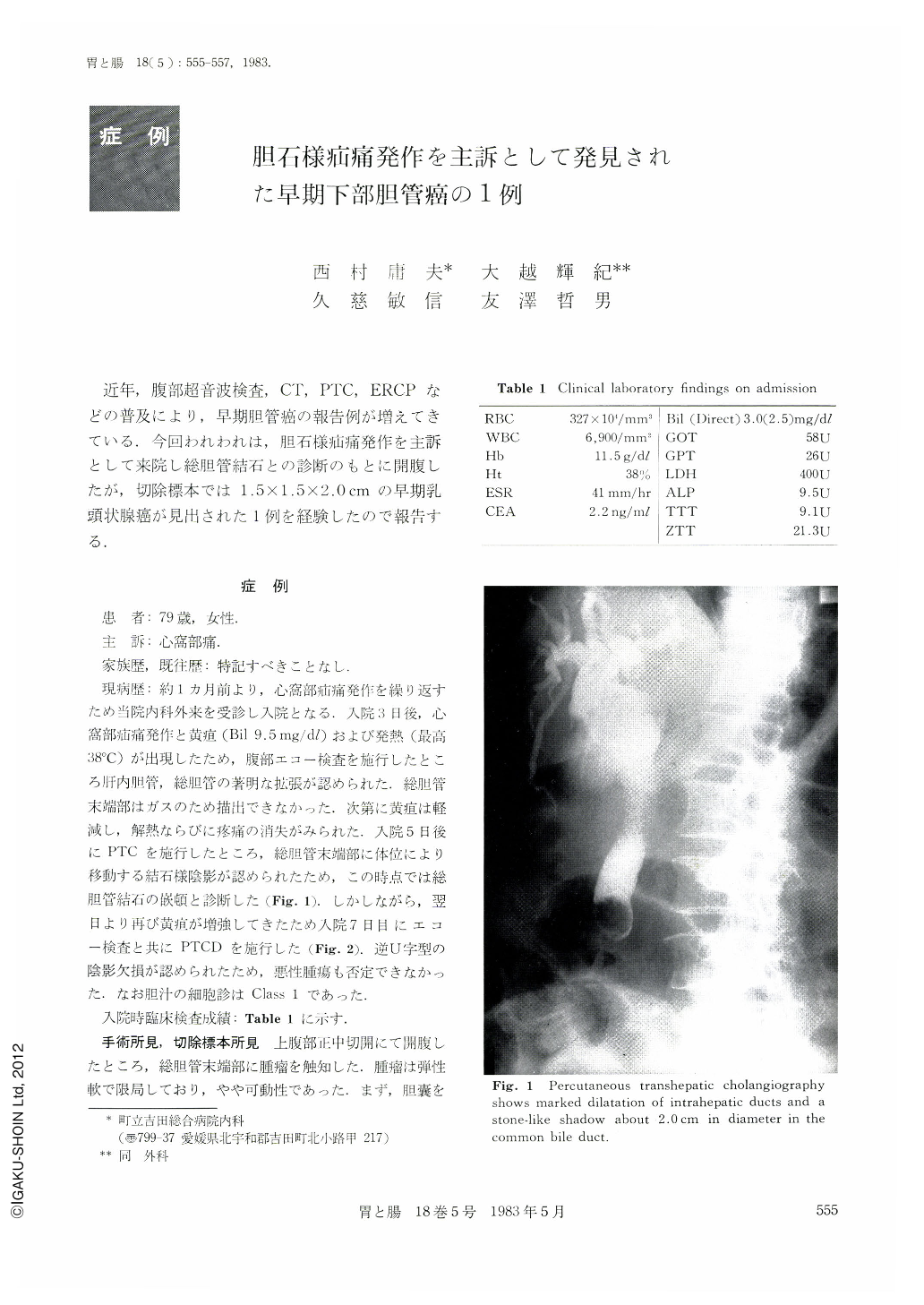
近年,腹部超音波検査,CT,PTC,ERCPなどの普及により,早期胆管癌の報告例が増えてきている.今回われわれは,胆石様疝痛発作を主訴として来院し総胆管結石との診断のもとに開腹したが,切除標本では1.5×1.5×2.0cmの早期乳頭状腺癌が見出された1例を経験したので報告する.
A 79 year-old woman patient was hospitalized with epigastric pain. Ultrasonic examination showed dilatation of the common bile duct. Percutaneous transhepatic cholangiography (PTC) showed marked dilatation of intrahepatic ducts and a stone-like shadow about 2.0 cm in diameter in the common bile duct. A few days later, because of marked stenosis of the common bile duct, PTC drainage was performed. As a result, a reversed U sign was seen in the common bile duct. Our preoperative diagnosis was a stone in the common bile duct. But, during the operation, we found a polypoid lesion in the common bile duct. Therefore, pancreatoduodenectomy was performed. Histologically diagnosis was papillary adenocarcinoma. About ten months have passed since the surgical operation, and the patient is in good health with no sign of recurrence.
Copyright © 1983, Igaku-Shoin Ltd. All rights reserved.