Japanese
English
- 有料閲覧
- Abstract 文献概要
- 1ページ目 Look Inside
- サイト内被引用 Cited by
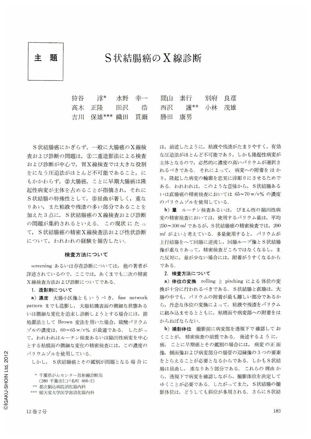
S状結腸癌にかぎらず,一般に大腸癌のX線検査および診断の問題は,①二重造影法による検査および診断が中心で,胃X線検査では大きな役割をになう圧迫法がほとんど不可能であること,にもかかわらず,②大腸癌,ことに早期大腸癌は隆起性病変が主体を占めることが指摘され,それにS状結腸の特殊性として,③屈曲が著しく,重なりあい,また粘液や残渣の多い部分であることを加えた3点に,S状結腸癌のX線検査および診断の問題が集約されるといえる.この現状にたって,S状結腸癌の精密X線検査法および性状診断について,われわれの経験を報告したい.
Ⅰ. Method of the Examination.
1) Concentration and an amount of the barium.
Since the early cancer of the sigmoid colon is protruded one and there are often found the mucus and the fecal sediments in the sigmoid colon, we usually use the barium with concentration of 65~70 w/v% and with an amount of the barium 200 ml for the double contrast roentgenography well known as one stage method. Brown's modification is best choice of premedication for the roentgenological examination of the colon.
2) Method of the examination.
It is very important to change well enough the position of the patient on taking the picture. In double contrast roentgenography, position of the patient is usually kept antero-posteriorly and right or left anteriorly on the supine position because of tortusity and overlapping of the loop of the sigmoid colon. Not only the method of compression, but also that of thin-barium coating are usefull for visualizing the lesions in detail.
Ⅱ. Diagnosis of the Cancer of the Sigmoid Colon.
In our series 23 lesions of 21 cases of the early cancer of the sigmoid colon, they are classified macroscopically into type Ⅰ, type Ⅱa and type Ⅱa+Ⅱc.
These are compared with 50 cases of the advanced cancer in each respects of size, depth of invasion and macroscopic features (Table 1,2,3 and 4), and the principles of diagnosis are summarized.
Thus, we can make an approach to diagnosis by the way as described as below;
1) Protruded type of the colonic lesions which are sessile and have a central depression is cancer. They are early cancer type Ⅱa+Ⅱc if small in size, but advanced one if large.
2) Since the early cancer type Ⅰ is pedunculated or subpedunculated, it is very often impossible to differentiate from the benign polyps.
It is neccessary to polypectomize them for the complete biopsy.
3) Diagnosis of type Ⅱa is possible because they are usually low in height, oval and irregular in shape.
4) Deformity of the intestinal wall.
Cancer of the sigmoid colon shows bilateral and unilateral deformity of the intestinal wall. Bilateral deformity of the wall is seen in case of the advanced cancers (Fig. 1).
Ⅲ. Case presentation.
Fig. 2 and Fig. 3: Case of advanced cancer (Borrmann 3) of the sigmoid colon.
The pictures which are taken much carefully and delibreate observation are demanded because even the advanced cancer is missed in the sigmoid colon.
Fig. 4 and Fig. 5: Case of the early cancer type Ⅱa+Ⅱc. In Fig. 4, the lesion is not shown on the routine roentgenography. Fig. 5 is picture of further examination. Lesion is revealed by combination of the double contrast radiography and compression. Fig. 6 and Fig. 7: Case of the early cancer type Ⅰ. In Fig. 7. the pedicle of the tumor is shown by compression.
Fig. 8 and Fig. 9: Case of the early cancer type Ⅰ.
Fig. 9 is picture by thin barium coating.
Fig. 10: Early cancer type Ⅰ. Fig. 11: Early cancer type Ⅰ. Fig. 12: Early cancer type Ⅱa. Fig. 13: Early cancer type Ⅱa+Ⅱc with central depression.

Copyright © 1977, Igaku-Shoin Ltd. All rights reserved.


