Japanese
English
- 有料閲覧
PPV購入案内
1,320円 (1,200円+税10%) こちらは、221ページから225ページの文献です。購入には医書.jp本会員登録が必要です。
会員登録完了後に改めて上記「購入サイトへ」を選択してください。
または「購入サイトへ」選択後に「購入ログイン」、「新規会員登録」の順に進んでください。
会員登録について詳しくはをご確認ください。
- Abstract 文献概要
- 1ページ目 Look Inside
小腸の形態学的診断法としては,従来から腹部単純X線検査にはじまり,X線透視,血管造影,string-test,腸紐,盲目的小腸生検などがその中心としてなされてきたが,その診断は困難なことが多く,臨床的にも原因不明の腸閉塞,腸管穿孔,下血等の重篤な症状を呈して,開腹術や剖検によってはじめて診断がなされることがたびたびである1)2).
一方,小腸の内視鏡観察の試みとして,経腹腸鏡による回盲部観察3)4),十二指腸fiberscopeによる空腸上部の観察,大腸 fiberscopeによる逆行性の回腸終末部の観察などが試みられてきたが5)~7),近年,より広範囲の小腸の内視鏡観察を目的として,小腸fiberscopeの試作・開発がさなれている4)~9).
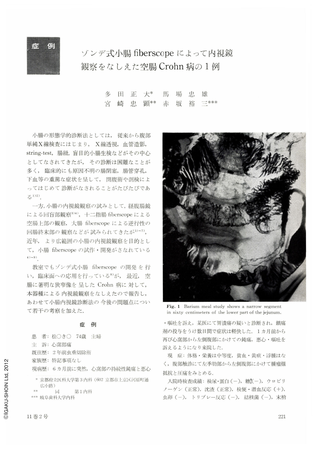
A 74 years old female was admitted to our hospital with six months' history of epigastric pain and vomiting. Barium meal study showed a narrowed segment 60 cm distant from the Treitz ligamentum of the jejunum. After that, small intestinal fiberscopy using specially designed “Sonde” type fiberscope (Olympus) revealed diffuse erosion, ulceration and reddish granular appearance of the mucosa. The resected specimen showed the same appearance as was observed by endoscopy, and its histological finding was granulomatous enteritis (Crohn's disease).
In Japan,Crohn's disease is rare and only 275 cases have been reported in recent 20 years, and only 7 (2.5%) of 275 cases originated in the jejunum. Its small intestinal fiberscopic observation has not been reported until now.
Copyright © 1976, Igaku-Shoin Ltd. All rights reserved.