Japanese
English
- 有料閲覧
PPV購入案内
1,320円 (1,200円+税10%) こちらは、647ページから651ページの文献です。購入には医書.jp本会員登録が必要です。
会員登録完了後に改めて上記「購入サイトへ」を選択してください。
または「購入サイトへ」選択後に「購入ログイン」、「新規会員登録」の順に進んでください。
会員登録について詳しくはをご確認ください。
- Abstract 文献概要
- 1ページ目 Look Inside
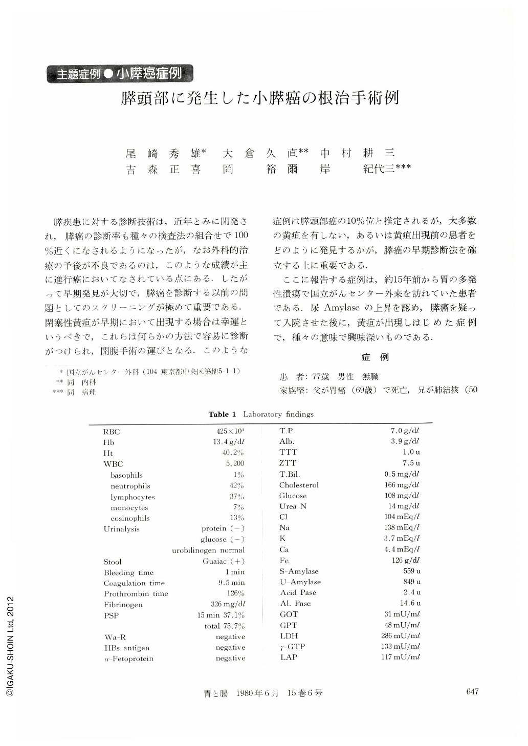
膵疾患に対する診断技術は,近年とみに開発され,膵癌の診断率も種々の検査法の組合せで100%近くになされるようになったが,なお外科的治療の予後が不良であるのは,このような成績が主に進行癌においてなされている点にある.したがって早期発見が大切で,膵癌を診断する以前の問題としてのスクリーニングが極めて重要である.閉塞性黄疸が早期において出現する場合は幸運というべきで,これらは何らかの方法で容易に診断がつけられ,開腹手術の運びとなる.このような症例は膵頭部癌の10%位と推定されるが,大多数の黄疸を有しない,あるいは黄疸出現前の患者をどのように発見するかが,膵癌の早期診断法を確立する上に重要である.
ここに報告する症例は,約15年前から胃の多発性潰瘍で国立がんセンター外来を訪れていた患者である.尿Amylaseの上昇を認め,膵癌を疑って入院させた後に,黄疸が出現しはじめた症例で,種々の意味で興味深いものである.
The patient was a 77 year-old male who had been followed up because of gastric ulcer since 1964. On February 16, 1979, he suddenly had migrane with anorexia and hoarseness of the voice. At that time glycosuria was detected by his home doctor. In April, abnormally high level of amylase was detected in his urine and serum and he was admitted to the National Cancer Center Hospital for further examinations.
Endoscopic retrograde cho1angio-pancreatography revealed a remarkable stenosis of the main pancreatic duct in the head of the pancreas, suggesting carcinoma of the pancreas. The caudal part of the pancreatic duct was tortuous and dilated. The dilatation of the caudal pancreatic duct was also med by computed tomography and ultrasonography. However, malignancy was not pointed out by these examinations. Selective abdominal angiography was negative for malignancy.
On June 19, 1979, the patient was operated on. Marked adhesion was observed in the upper abdominal cavity, but neither ascites nor metastatic lesions were detected. A pigeon-egg sized tumor was found in the head of the pancreas. On palpation the caudal part of the pancreas was hard and there was possibility of cancer infiltration to the pancreatic body and tail, so total pancreatectomy was carried out.
On cut surface of the resected specimen, a tumor measuring 1.8×1.2 cm was observed in the head of the pancreas. The main pancreatic duct adjoining the tumor was stenotic and the caudal part was remarkably dilated. Histological diagnosis was moderately differentiated adenocarcinorna. Five of the 57 lymph nodes examined were positive for cancer cells.
The patient is alive at this writing. (May, 1980)
Copyright © 1980, Igaku-Shoin Ltd. All rights reserved.