Japanese
English
- 有料閲覧
PPV購入案内
1,320円 (1,200円+税10%) こちらは、771ページから775ページの文献です。購入には医書.jp本会員登録が必要です。
会員登録完了後に改めて上記「購入サイトへ」を選択してください。
または「購入サイトへ」選択後に「購入ログイン」、「新規会員登録」の順に進んでください。
会員登録について詳しくはをご確認ください。
- Abstract 文献概要
- 1ページ目 Look Inside
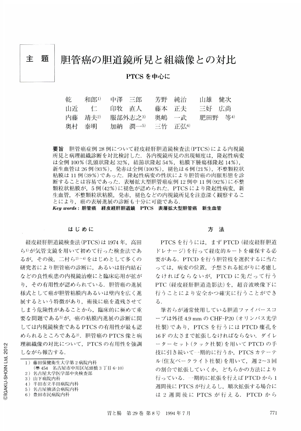
要旨 胆管癌症例28例について経皮経肝胆道鏡検査法(PTCS)による内視鏡所見と病理組織診断を対比検討した.各内視鏡所見の出現頻度は,隆起性病変は全例100%(乳頭状隆起32%,結節状隆起54%,粘膜下腫瘍様隆起14%),新生血管は26例(93%),発赤は全例(100%),褪色は6例(21%),不整顆粒状粘膜は11例(39%)であった.隆起性病変の性状により胆管癌の肉眼形態を診断することは容易であった.表層拡大型胆管癌症例12例中11例(92%)に不整顆粒状粘膜が,5例(42%)に褪色が認められた.PTCSにより隆起性病変,新生血管,不整顆粒状粘膜,発赤,褪色などの内視鏡所見を注意深く観察することにより,癌の表層進展の診断も十分に可能である.
We reported endoscopic findings with percutaneous transhepatic cholangioscopy and pathological findings in bile duct cancer. In 28 patients with bile duct cancer, protrusion was observed in 100% of the cases, neoplastic vascularity in 93%, redness in 100%, discoloration in 21%, and irregular granular mucosa in 39%. We were able to classify "protrusion" as papillary, nodular and submucosal-like. We were able also to verify and distinguish these features grossly in resected specimens. In 12 cases of superficial-spreading cancer diagnosed correctly with percutaneous transhepatic cholangioscopy, irregular granular mucosa was observed in 92% and discoloration was observed in 42%.
Copyright © 1994, Igaku-Shoin Ltd. All rights reserved.