Japanese
English
- 有料閲覧
PPV購入案内
1,320円 (1,200円+税10%) こちらは、653ページから659ページの文献です。購入には医書.jp本会員登録が必要です。
会員登録完了後に改めて上記「購入サイトへ」を選択してください。
または「購入サイトへ」選択後に「購入ログイン」、「新規会員登録」の順に進んでください。
会員登録について詳しくはをご確認ください。
- Abstract 文献概要
- 1ページ目 Look Inside
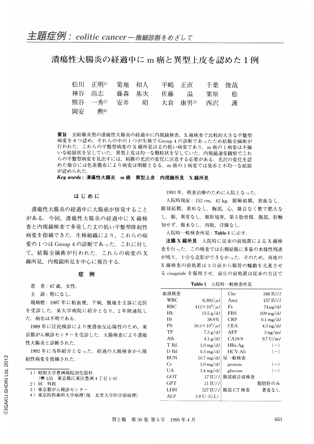
要旨 全結腸炎型の潰瘍性大腸炎の経過中に内視鏡検査,X線検査で比較的大きな平盤型病変を8つ認め,それらの中の1つが生検でGroup4の診断であったため結腸全摘術が行われた.これらの平盤型病変のX線所見は丈の低い病変であり,m癌の1病変は不揃いな結節状を呈していた.異型上皮は均一な顆粒状を呈していた.内視鏡通常観察でこれらの平盤型病変を見出すには,粘膜の光沢の変化に注意する必要がある.光沢の変化を認めた場合には色素撒布により病変は明瞭となる.m癌の1病変では発赤と不均一な結節が認められた.
A 67-year-old female, who had had ulcerative colitis (total colitis) since the age of 61, was examined by endoscopy and barium enema because of bloody stool and diarrhea. The endoscopic examination revealed eight plaque-like lesions in the large bowel. In one of them histological examinations of the biopsy recognized Group 4. Radiological findings of plaque-like lesion were demonstrated as radiolucent lesions and endoscopic findings, using a dye-spray method, were plaque-like lesions with a nodular pattern.
Total colectomy with ileo-rectal anastomosis was performed. The histological examination showed that two mucosal cancers of the cecum and transverse colon were well differentiated adenocarcinomas and six plaque-like lesions of the colon ascendens, transverse and descendens were dysplasia, and that colonic mucosa was in a remission stage.
Copyright © 1995, Igaku-Shoin Ltd. All rights reserved.